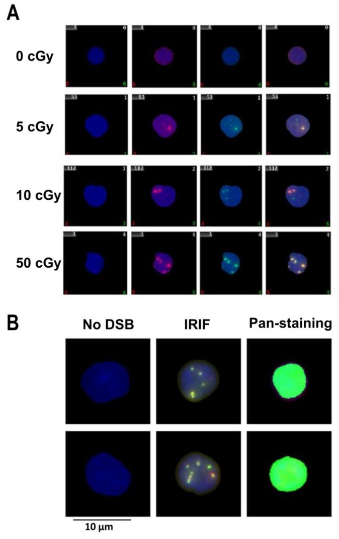
Figure 1.
Representative images of nuclei from human UCB. Representative images of: (A) the nuclei (stained with DAPI in blue) from the human cord blood lymphocytes 30 min post-irradiation with 0 (sham irradiated control), 5, 10, and 50 cGy: γH2AX foci (green), 53BP1 (red), co-localized γH2AX/53BP1 overlay of green and red (yellow); and (B) representative raw images of pan-nuclear staining of γH2AX observed in human cord blood lymphocytes 22 h post-irradiation by the dose of 50 cGy.

