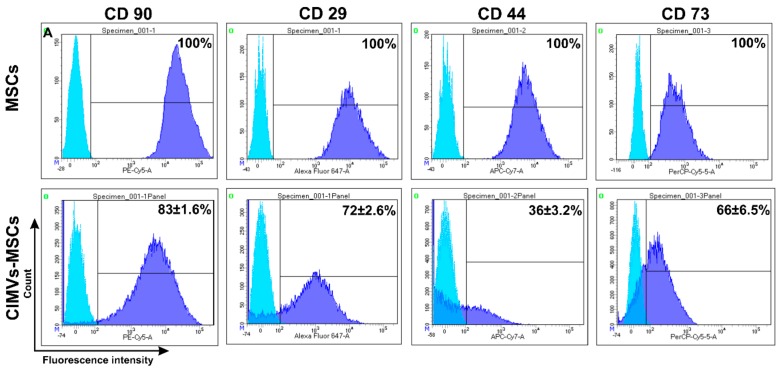
Figure 4.
Immune phenotype of human MSCs and CIMVs-MSCs. MSCs and CIMVs-MSCs were stained with anti-CD90, anti-CD29, anti-CD44, and anti-CD73 monoclonal antibodies and analyzed using flow cytometer BD FACS Aria III (BD Bioscience, San Jose, CA, USA). Histograms were generated using the FACSDiva7 software (BDBioscience, Version 7.0, San Jose, CA, USA). Blue—isotype control; dark blue—MSCs or CIMVs-MSCs labeled with antibodies.

