Figure 5.
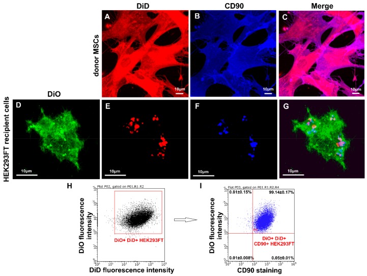
Analysis of CD90 transfer by CIMVs-MSCs to recipient HEK293FT cells. (A–G) Laser scanning confocal microscopy using Zeiss LSM 780 (Carl Zeiss, Oberkochen, Germany), (H–I) flow cytometry using BD FACS Aria III (BD Bioscience, San Jose, CA, USA). Green fluorescence—recipient HEK293 FT cell stained with DiO; red fluorescence—parental MSCs or CIMVs-MSCs stained with DiD, blue fluorescence—cells stained with anti-CD90 antibody. (A–C) MSCs stained with DiD and anti-CD90 antibody. (D–G) HEK293FT cells treated with CIMVs-MSCs (CIMVs-MSCs—red spots) and stained with DiO and anti-CD90 antibody.

