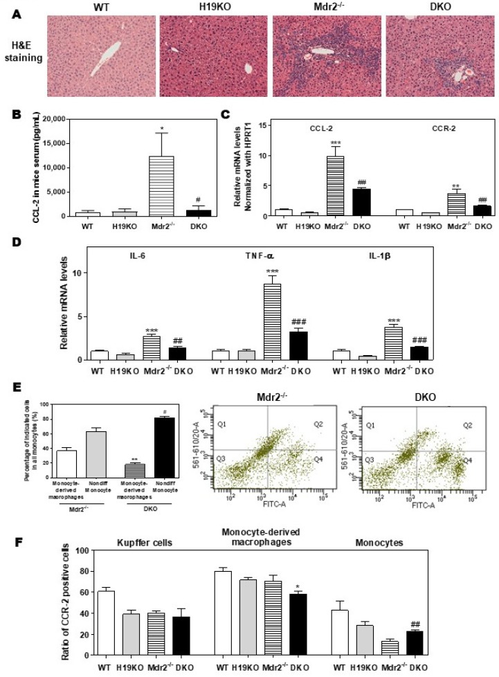
Figure 5.
H19-enriched exosomes promote liver inflammation and macrophage activation in Mdr2-/- mice. WT, H19KO, Mdr2-/- mice, and DKO mice (both male and female at 100-day old) were sacrificed. (A) Representative images of H&E staining are shown. (B) CCL-2 levels in the serum. (C,D) The relative mRNA levels of hepatic CCL-2, CCR-2, IL-6, TNF-α, and IL-1β were determined by real-time RT-PCR and normalized using HPRT1. Statistical significance: * p < 0.05, ** p < 0.01, *** p < 0.001, compared with WT mice; # p < 0.05, ## p < 0.01, ### p < 0.001, compared with Mdr2-/- mice. (E,F) Cell type-specific markers were used to determine the cell population using flow cytometry analysis. (E) Representative flow cytometry results and images of the percentage of indicated cells in all monocytes are shown. (F) The ratio of CCR-2 (C-C chemokine receptor 2) positive cells in Kupffer cells, monocyte-derived macrophages, and monocytes. Statistical significance: * p < 0.05, ** p < 0.01, compared with monocyte-derived macrophages in Mdr2-/- mice; # p < 0.05, ## p < 0.01, compared with non-differentiated monocytes in Mdr2-/- mice (n > 6).

