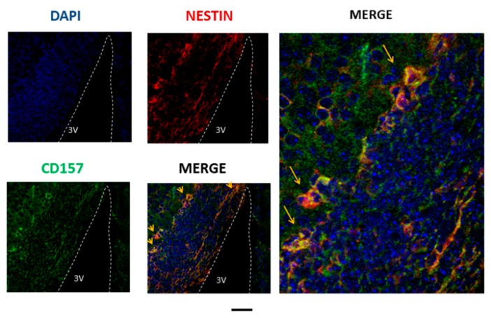
Figure 1.
Cluster of differentiation 157 (CD157) expression analyzed by immunofluorescence staining in the embryonic mouse brain. Representative images were obtained from the E17 embryonic hypothalamus near the third ventricle (3V). CD157 is stained in green, nestin in red, and the nucleus in blue. Two merged images show the expression of CD157 in neural stem cells. Scale bar, 100 and 30 μm for the four left and enlarged images, respectively.

