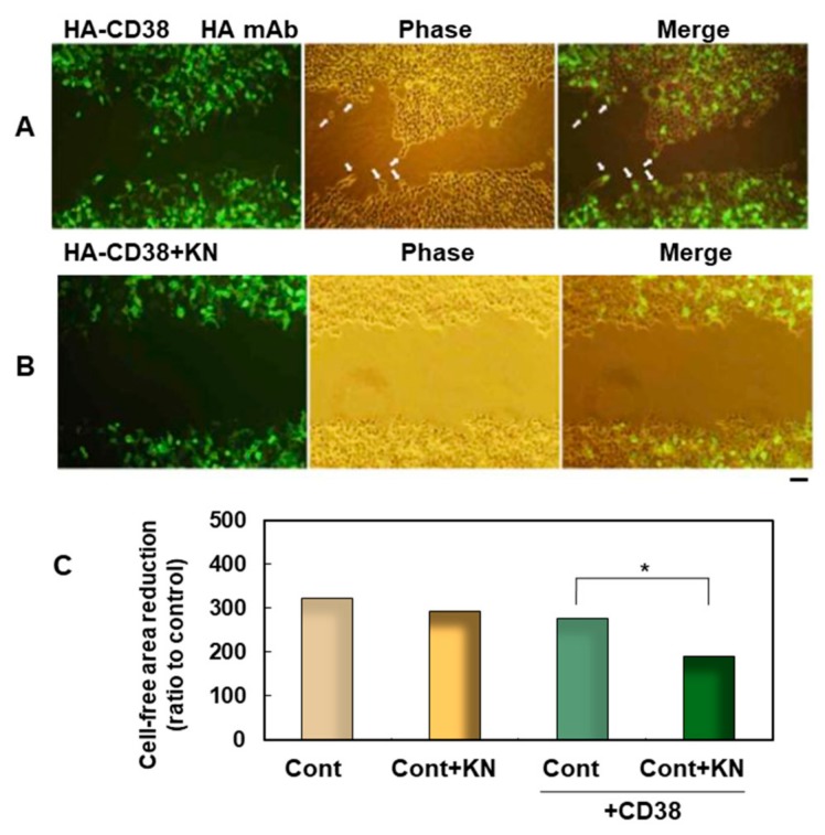
Figure 3.
Effect of a CAMKII inhibitor on wound-induced migration facilitated by the expression of human influenza hemagglutinin (HA)-hCD38. (A,B) Human embryonic kidney 293T (HEK293T) cells grown on plastic dishes were transfected with HA-hCD38 complementary DNA (cDNA) constructs. One day after transfection, a cell-free area was formed by scratching the plastic dishes, and they were treated with 0.1% dimethyl sulfoxide (DMSO) solution (control, A) or with 1 µM KN-62, a CaM kinase inhibitor, dissolved in 0.1% DMSO solution (B). At 24 h after scratching, they were fixed, permeabilized, and stained with HA monoclonal antibody (mAb). The right panels are merged images of fluorescent (Left) and phase-contrast (Middle) pictures. Scale bar, 100 µm. (C) Quantification of cell migration using the monolayer wound healing assay. The degree of migration was expressed as a reduction of the cell-free area by occupied cells. Each bar corresponds to 293T cells transfected with (green bars) or without (yellow bars) 1 µM of KN-62 (+KN). Each bar is the mean ± standard error of the mean (SEM) in seven experiments. Two-way ANOVA, F3,7 = 7.98, p = 0.0001. A significant difference was seen in the conditions of CD38 transfection and KN treatment, but no significant interaction between transfection of CD38 and treatment with KN-62 was observed (p = 0.18). Bonferroni’s post hoc analysis revealed a significant difference from the KN-free control value (*p < 0.05).

