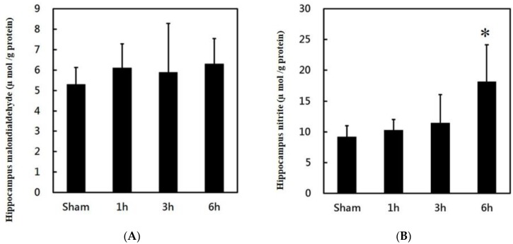
Figure 6.
Effects of PPTg DBS on hippocampal oxidative stress: (A) illustration of oxidative stress marker malondialdehyde (MDA) and (B) illustration of nitrite levels in hippocampal tissue was determined at hours 1, 3, and 6-time intervals. Data were presented as mean ± SD (n = 6). Significant differences between groups were analyzed using one-way ANOVA. * p < 0.05 was considered statistically significant compared with the sham group.

