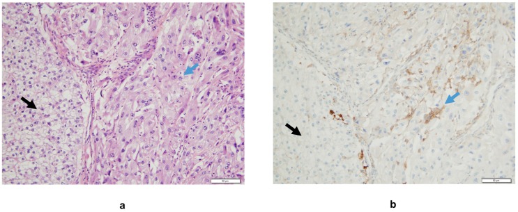
Figure 2.
(a) Interface between areas of classical clear-cell carcinoma (black arrow) and its sarcomatoid component (blue arrow), Hematoxylin Eosin staining. (b) A higher density of tumor-infiltrating mononuclear inflammatory cells expressing PD-L1 is observed in the sRCC area compared to the clear-cell RCC one (monoclonal mouse antibody clone 22C3, hematoxylin counter staining). In this case, there is no PD-L1 expression on tumor cells.

