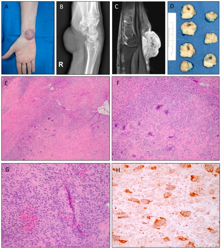
Figure 1.
Clinical presentation, radiologic features, and histomorphology of Case 1. (A) A soft tissue mass was seen located on the volar aspect of the wrist. (B) Lateral X-ray of the right wrist revealed a nonspecific mass. (C) Sagittal T1 fat-suppressed postcontrast MRI showed hyperintensity of the lesion. (D) Gross appearance of the excised tumor demonstrated an area of central hemorrhage encased by a partial deep fibrous band. (E) Photomicrograph with low-power magnification showing a cellular neoplasm growing in a vague fascicular pattern with scattered osteoclast-like multinucleated giant cells and areas of hyaline matrix deposition and focal osteoid matrix formation (original magnification ×4). (F) Areas of hypercellularity with stromal hyalinization and osteoid deposition (original magnification ×10). (G) Higher-power magnification demonstrated areas of increased cellularity with spindle cells with enlarged nuclei, open chromatin, and focal enlarged nucleoli. Note the lack of necrosis (original magnification ×20). (H) Tumor cells displayed immunopositivity for CD68, both mononuclear and multinucleated cells.

