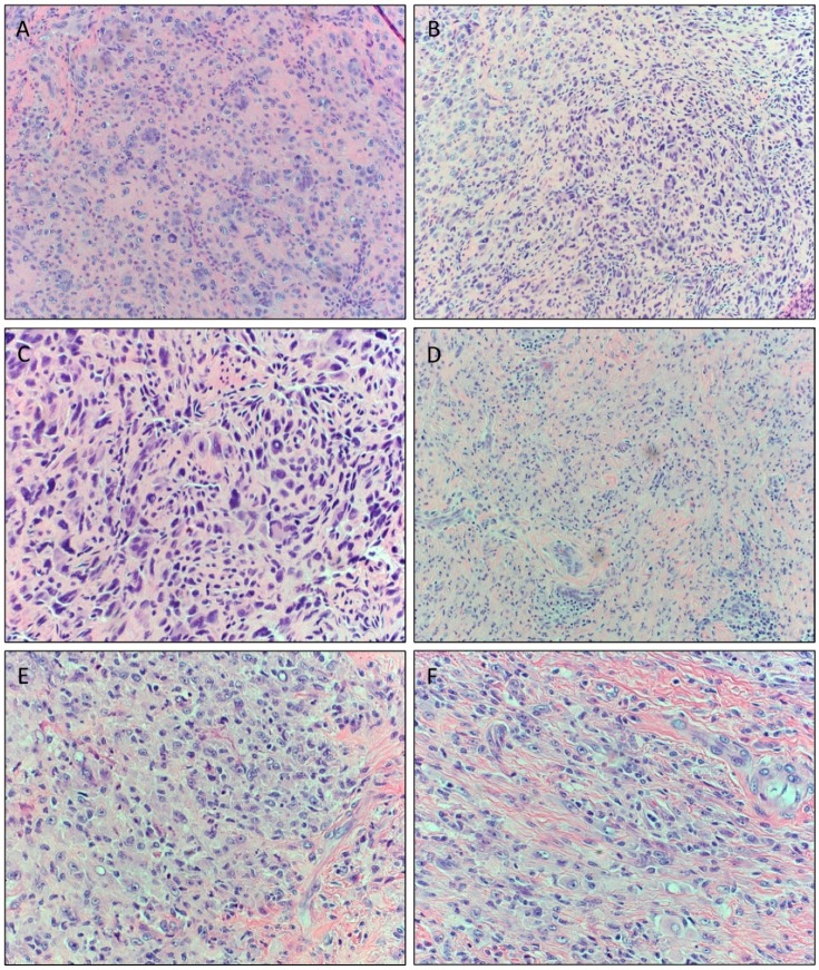
Figure 2.
Spectrum of histomorphologic features of atypical tenosynovial giant cell tumors (TGCTs) in Case 2 (A–C) and Case 3 (D–F). (A) Mid-power magnification showing a cellular neoplasm with a predominance of mononucleated cells with scattered multinucleated osteoclast-like cells with intervening fibrous stroma (original magnification ×20). (B) Areas showing hyperchromatic pleomorphic nuclei and spindling. No necrosis was seen (original magnification ×40). (C) High-power magnification demonstrating multinucleated giant cells with symplastic changes in addition to increased cellular atypia in the mononuclear cell population consistent with the rendered diagnosis of atypical component within the tumor (original magnification ×40). (D) Photomicrograph of more benign-appearing areas composed of multinucleated giant cells and sheets of epithelioid histiocytoid cells arranged in a syncytial pattern (original magnification ×20). (E) Areas showing presence of larger pleomorphic nuclei with open chromatin and prominent nucleoli (original magnification ×40). (F) Scattered areas of cells with enlarged nuclei, spindle cells, and cells with virocyte-like nucleoli conferring worrisome morphology (original magnification ×40).

