Figure 2.
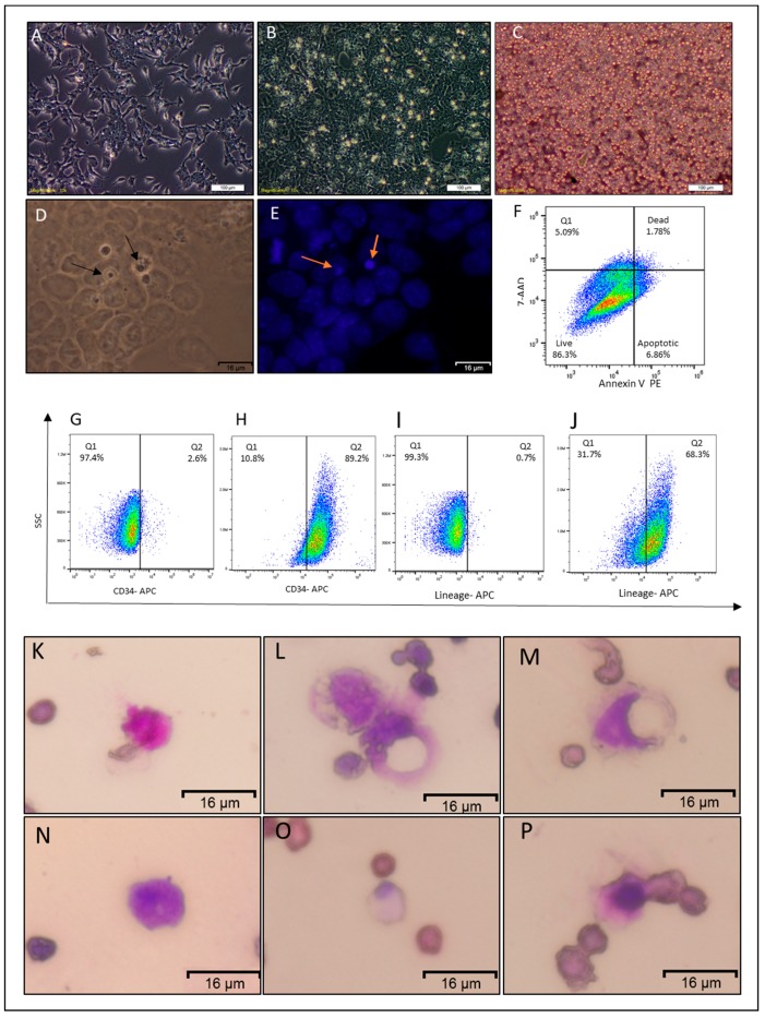
Characterization of the non-adherent round cells. (A) Representative image of CSCcmBT549 after 24 h of seeding. (B) Representative images of CSCcmBT549 cells after 72 h of seeding, showing round non-adherent cells on the top of the monolayer of adherent cells. (C) Floating non-adherent cells collected from the culture of CSCcmBT549 cells. Scale bars for (A,B,C) represent 100 μm. (D,E) Bright field and DAPI staining showing nuclei of round non-adherent cells (NACs) on the top of the monolayer adherent cells. Scale bars represent 16 μm. (F) Representative image of flow cytometry analysis of apoptosis assay by” Annexin V and 7-AAD kit” shows that the majority of the cells are viable while apoptotic and dead cells are less than 15%. This image is representative of at least three independent experiments. (G–J) Flow cytometry analysis for CD34 and hematopoietic lineage differentiation markers (Lineage Cell Detection Cocktail-Biotin, where (G,I) are for adherent CSCcmBT549 cells and (H,J) are for NACs. Each result is shown as a representative of at least three independent experiments. (K–P) Wright–Giemsa staining of floating cells showing different diameters and staining patterns. Scale bars represent 16 μm.

