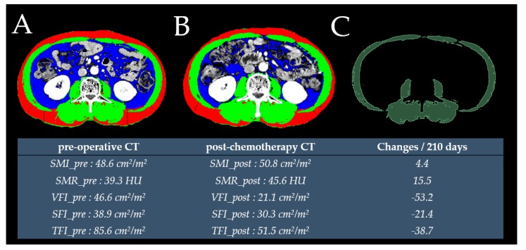
Figure 2.
Cross-sectional L3 CT image used to measure subcutaneous fat (red), skeletal muscle (green) and visceral fat (blue) using preoperative (A) and postchemotherapy CT (B) images. The radiodensity of skeletal muscle in the preoperative CT was measured using an open-source three-dimensional slicer software (C).

