Figure 2.
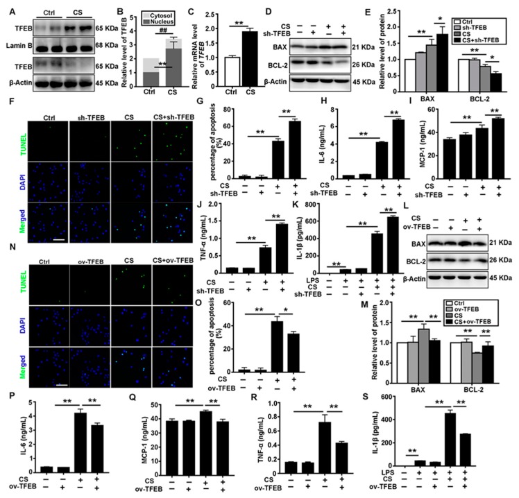
TFEB modulates CS-induced macrophage apoptosis and inflammatory response in vitro. (A,B) Immunoblotting analysis of TFEB levels in the cytosol and nucleus of MH-S cells 12 h post saline or CS treatment; β-actin, loading control for cytosolic proteins and lamin B, loading control for nuclear proteins (n = 4). (C) qPCR analysis of TFEB expression in MH-S cells 12 h post-saline or CS treatment (n = 4). (D,E) TFEB knockdown deteriorates CS-induced apoptosis and inflammation. Immunoblotting analysis of BAX and BCL-2 protein levels (n = 3 to 4). (F,G) TUNEL (green) and DAPI (blue) staining and ratios of TUNEL-positive apoptotic cells (scale bar = 50 μm and n = 4). (H–K) ELISA analysis of IL-6, MCP-1, TNF-α, and IL-1β levels in MH-S cell supernatant (n = 3 to 4). (L,M) TFEB overexpression relieves CS-induced apoptosis and inflammation. Immunoblotting analysis of BAX and BCL-2 protein levels (n = 4). (N,O) TUNEL (green) and DAPI (blue) staining and ratios of TUNEL-positive apoptotic cells (scale bar = 50 μm and n = 4). (P–S) ELISA analysis of IL-6, MCP-1, TNF-α, and IL-1β levels in MH-S cell supernatant (n = 3 to 4). Data are presented as the mean ± SD. *, p < 0.05; **, p < 0.01; and ##, p < 0.01.

