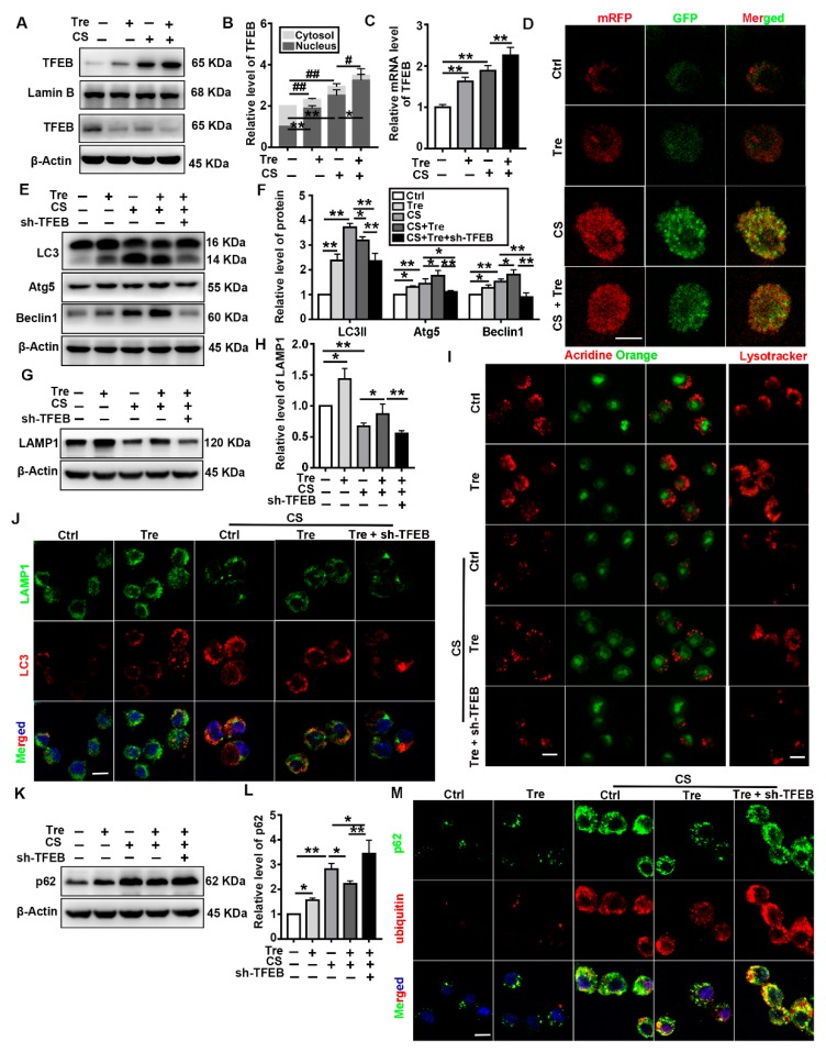
Figure 6.
Tre relieves CS-induced lysosome damage and restores autophagic substrate degradation through TFEB activation in vitro. (A,B) Immunoblotting analysis of TFEB levels in the cytosol and nucleus of MH-S cells post CS and Tre treatment and β-actin, loading control for cytosolic proteins; LaminB, loading control for nuclear proteins (n = 3 to 4). (C) qPCR analysis of TFEB expression in MH-S cells 12 h post-CS and Tre treatment (n = 3 to 4). (D) MH-S cells were transfected with the adenovirus mRFP-GFP-LC3 plasmid. LC3 puncta were observed by confocal microscopy (scale bar = 10 μm and n = 3). (E,F) Immunoblotting analysis of LC3II, Atg5, and Beclin 1 protein levels (n = 3). (G,H) Immunoblotting analysis of LAMP1 protein levels in cell lysates (n = 4). (I) Cells were grown on coverslips and stained with Lysotracker Red or acridine orange 12 h post CS and Tre treatment (scale bar = 10 μm and n = 3). (J) Immunofluorescence analysis of the colocalization of LAMP1 and LC3 12 h post CS and Tre treatment (scale bar = 25 μ and n = 3). (K–M) Immunoblotting analysis of p62 and immunofluorescence analysis of the colocalization of p62 and ubiquitin 12 h post CS and Tre treatment (scale bar = 25 μm and n = 3). Data are presented as the mean ± SD. *, p < 0.05; **, p < 0.01; #, p < 0.05; and ##, p < 0.01.

