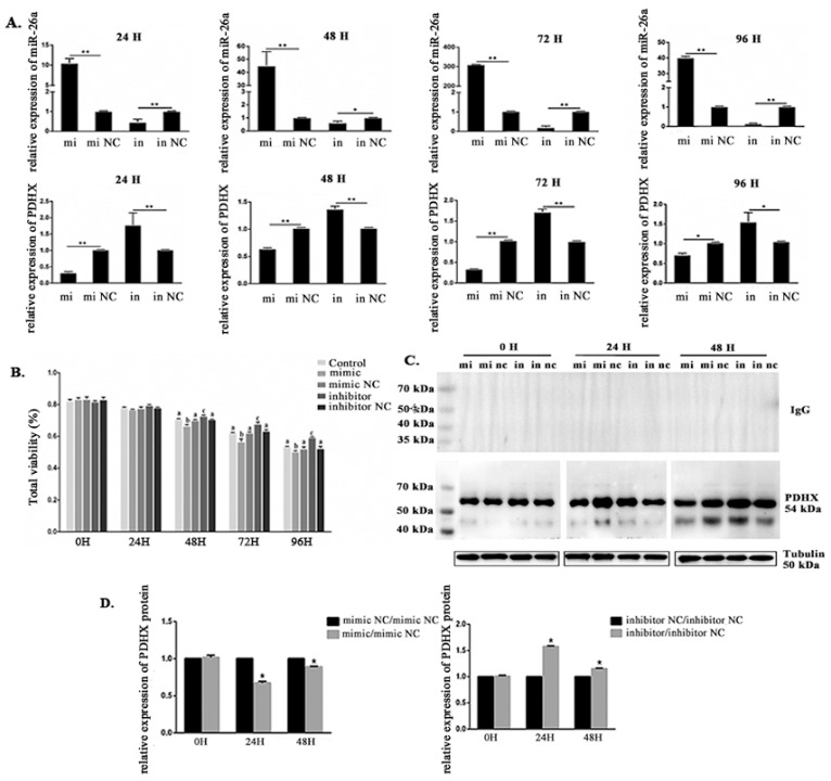
Figure 5.
The expression level of miR-26a and PDHX, and sperm viability in miR-26a transfected boar sperm at 17 °C. (A) Sperm viability after miR-26a transfection. (B) The expression level of miR-26a and PDHX after transfection with miR-26a mimic, mimic control, miR-26a inhibitor and inhibitor control for 24 h, 48 h, 72 h and 96 h, respectively. (C) WB analysis after 0 h, 24 h, and 48 h treatment with miR-26a mimic/mimic NC and inhibitor/inhibitor NC showing the expression of PDHX and β-tubulin. The negative control rabbit IgG in place of the primary antibody was used. (D) The relative protein level of PDHX after transfection with miR-26a mimic and inhibitor. Values were normalized using β-tubulin protein as an internal reference, and then mimic NC and inhibitor NC groups were used as references to measure the relative expression levels. mi: mimic; mi nc: mimic NC; in: inhibitor; in nc: inhibitor NC. “*” indicates statistical significance at P < 0.05 and “**” indicates statistical significance at P < 0.01. Different alphabets indicate statistical significance at P < 0.05.

