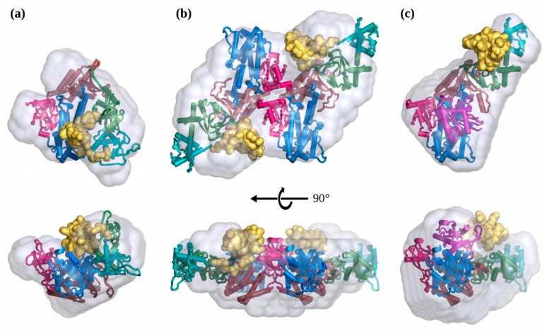
Figure 4.
Rigid-body SAXS Models of EPAC1 in each state. (a) Solution structure of apo-EPAC1, in its compact state, coloured by domain as in Figure 1. (b) Solution structure of the cAMP bound- EPAC1 dimer, looking down the two-fold axis. (c) Solution structure of the EPAC1:cAMP:Rap1b ternary complex. The same domain colour scheme is used for all figures unless indicated otherwise. The NTD is displayed as a solid yellow surface, and the DAMMIF molecular shape is displayed as a semi-transparent gray surface. The catalytic GEF (blue), RA (brown), and REM (red) domains are in the same orientation for each model. The CORAL model SAXS curve fits are shown in Figure 3 and Table 1.

