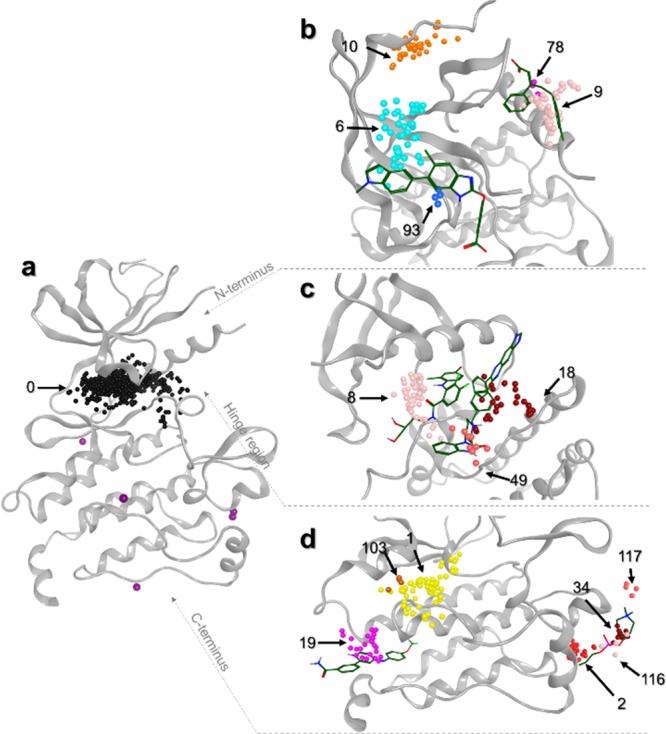
Figure 3.

Representation of heterogeneous clusters on a kinase structure (PDB ID 1K5V). (a) Orthosteric site represented by cluster 0 in black. Density points are still present in purple. (b) Heterogeneous clusters are displayed on the N-terminal region of the protein kinase. Reference ligands bound to AMPK (PDB ID 4CFE, left) and PDK1 (PDB ID 3HRF, right) are represented in sticks. (c) Heterogeneous clusters are displayed on the hinge region. Reference ligands bound to MEK1 (PDB ID 3EQC, left) and AKT1 (PDB ID 3O96, right) are represented in sticks. (d) Heterogeneous clusters are displayed on the C-terminal part. Reference ligands bound to ABL1 (PDB ID 3K5V, left) and MAPK14 (PDB ID 4E6C, right) are represented in sticks. Cluster IDs are indicated, and centroids are color-coded based on their cluster IDs.
