Figure 9.
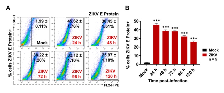
ZIKV (MOI 1) infects human endothelial (HMEC-1) cells. (A) ZIKV envelope (E) protein detection at 24, 48, 72, 96, and 120 h PI by the FACS assay. Dot plots are the representative mean ± SD of the positive cells from five independent experiments. (B) ZIKV-infected cells percentages obtained by FACS. The ZIKV E protein levels were compared (by an unpaired Student’s t-test) with the mock C6/36 (*) value. Statistical significance was recognized as * when p < 0.05, ** when p < 0.01, and *** when p < 0.0001.

