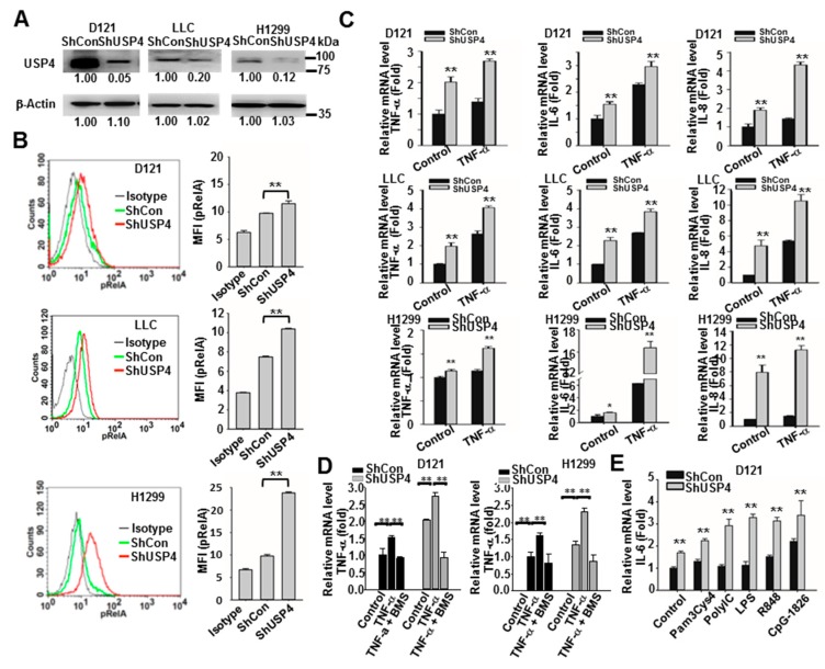
Figure 6.
Stable USP4 knockdown increases inflammatory status in lung cancer cells. (A) Efficiency of USP4 knockdown in lung cancer cell lines analyzed by immunoblotting. (B) NF-κB activation in control and USP4 knockdown lung cancer cells as measured by flow cytometric analysis of RelA phosphorylation. Left panels show representative histograms and right panels shows the quantitation of results. (C–E) Expression of cytokines in control and USP4 knockdown lung cancer cells stimulated with 10 ng/mL TNF-α with/without 1 μM BMS345541 (C,D) or 0.2 μg/mL Pam3Cys4, 5 μg/mL polyI:C, 0.2 μg/mL LPS, 2 μM R847, or CpG-1826 (E) for 24 h as analyzed by RT-qPCR. Data presented as mean ± SD of three independent experiments. * P < 0.05; ** P < 0.01.

