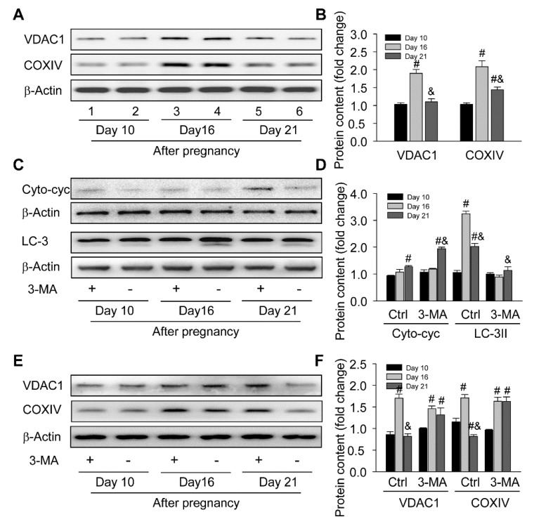
Figure 8.
Requirement of autophagy for mitochondria homeostasis in ovaries during the luteal development of pregnant rats: (A) representative western blot analyses depicting the protein levels of VDAC1 and COXIV in ovaries during the luteal development of pregnant rats; (B) summarized intensities of VDAC1 and COXIV blots normalized to the control; (C) representative western blot analyses depicting the protein levels of Cyto-cyc and LC-3 in ovaries with or without 3-MA treatment during the luteal development of pregnant rats; (D) summarized intensities of Cyto-cyc and LC-3II blots normalized to the control; (E) representative western blot analyses depicting the protein levels of VDAC1 and COXIV in ovaries with or without 3-MA treatment during the luteal development of pregnant rats; (F) summarized intensities of VDAC1 and COXIV blots normalized to the control. Each value represents the mean ± SE, n = 6. ANOVA was used to analyze the data. #: p < 0.05 vs. p on day 10 &: p < 0.05 vs. p on day 16. 3-MA: an autophagy inhibitor.

