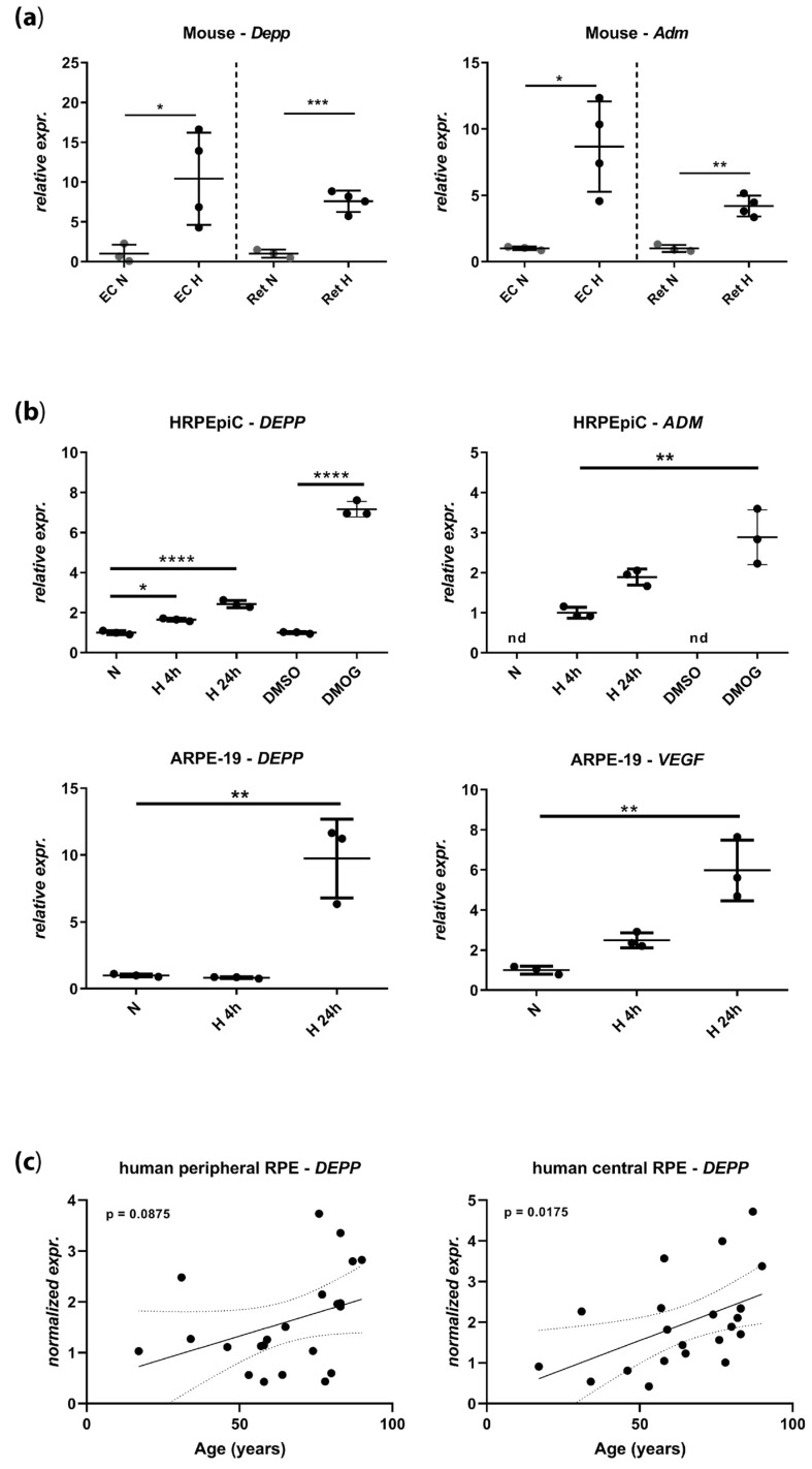
Figure 1.
Expression levels of decidual protein induced by progesterone (DEPP) and known hypoxia-inducible factors (HIF) target genes by semi-quantitative real-time PCR in (a) mouse eyecups (EC) and retina (normoxia: N = 3 eyes of three mice; hypoxia: N = 4 eyes of three mice) and (b) human retinal pigment epithelial cells (HRPEpiC) and human RPE cell line ARPE-19 cells. Levels in cells exposed to hypoxia (H) and cells treated with dimethyloxalylglycine (DMOG) are shown as fold changes over normoxic (N) or DMSO controls, respectively. Note that the transcript levels of adrenomedullin (ADM) in HRPEpiC are expressed relatively to the levels of 4 h of hypoxia. (c) DEPP transcript levels determined by semi-quantitative real-time PCR in human retinal pigment epithelium (RPE) samples collected from central and peripheral areas of donor eyes. Donor age ranged from 17 to 90 years (Table 1; N = 22). EC: Eyecup, Ret: Retina, Adm: Adrenomedullin, VEGF: Vascular endothelial growth factor, N: Normoxia, H: Hypoxia, nd: Not detected. Shown are individual values and means ± SD, Statistics: (A) and (B) one-way ANOVA with the Brown–Forsythe test, (C) linear regression. Dotted line indicates the p-value field. *: p ≤ 0.05, **: p ≤ 0.005, ***: p ≤ 0.005, ****: p ≤ 0.0005.

