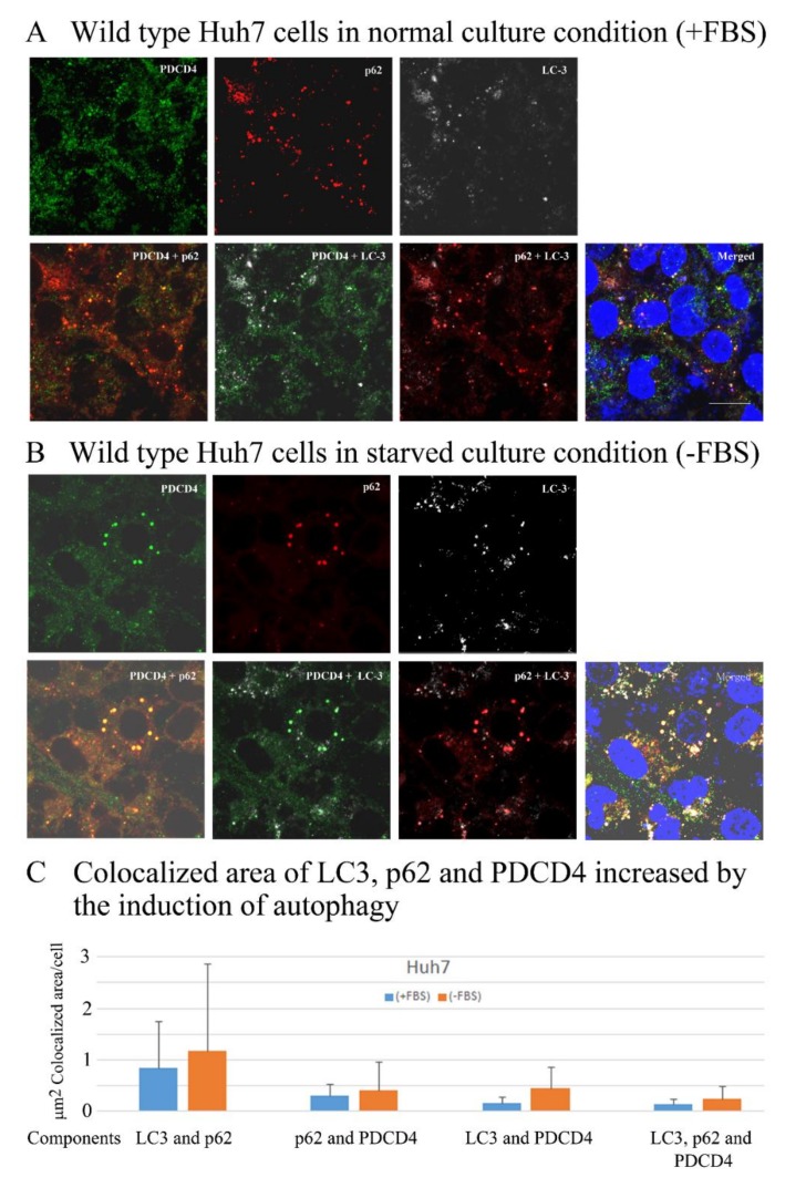
Figure 7.
PDCD4, p62. and LC3 were colocalized, and the colocalization areas expanded under serum-deprived conditions in wild-type Huh7 cells. (A,B) A total of 1.5 × 10⁵ cells were seeded onto a cover glass in 35-mm dishes and cultured until 70–80% confluency. The old medium was replaced with Dulbecco’s modified Eagle’s medium (DMEM) or DMEM + 10% FBS. The dishes were washed twice with DMEM before adding the new medium. Culture was continued for 4 h, and the cells were fixed with 4% paraformaldehyde. Immunostaining of the cells were performed as described in methods. Images were captured using an LSM-880 confocal microscope. The colocalization of PDCD4, p62, and LC3 in the presence (A) and absence (B) of FBS is shown. (C) A diagram of the colocalization areas of PDCD4, p62, and LC3 in wild-type Huh7 cells. Bar = 300 µm.

