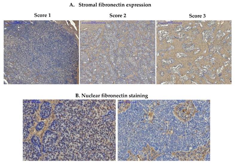
Figure 1.
Immunohistochemical detection of fibronectin in ovarian cancer samples. (A) Images show representative examples of stromal fibronectin staining, scored as 1 (weak expression), 2 (moderate expression), and 3 (strong expression); (B) Images show a representative example of nuclear staining in cancer cells (image on the left) and lack of nuclear staining in cancer cells (image on the right); Pannoramic 250 Flash II Scanner, scale bar: 500 µm (upper panel) and 100 µm (lower panel).

