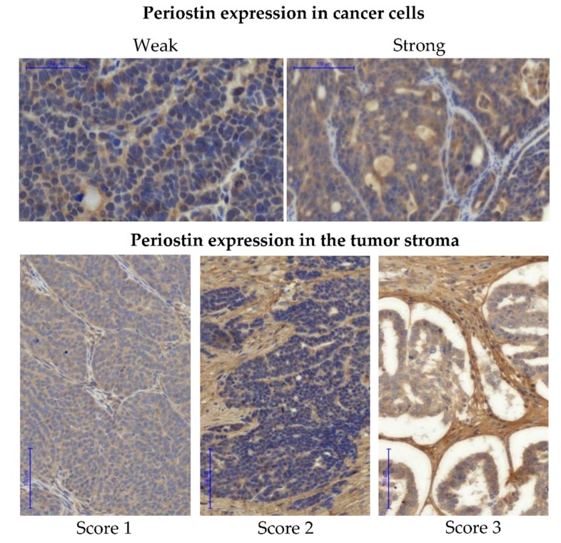
Figure 5.
Immunohistochemical detection of periostin. Periostin expression was evaluated separately in cancer cells (upper panel) and in the tumor stroma (lower panel). Images show IHC staining in cancer cells scored either as “weak” or “strong” and IHC staining of stromal periostin scored 1–3. Pannoramic 250 Flash II Scanner, scale bar: 50 µm and 100 µm.

