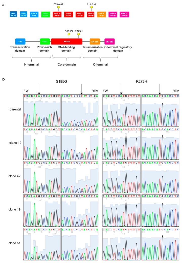
Figure 4.
PT-res MDAH clones gain a new TP53 missense mutation. (a) Schematic diagram of p53 gene (top) and protein (bottom). Exons are color-coded according to the corresponding protein functional domains (transactivation domain in blue, proline-rich domain in green, DNA-binding domain in red, tetramerization domain in orange, and C-terminal regulatory domain in pink). Yellow dots depict the mutations detected in MDAH PT-res cells. (b) Representative four-color fluorescence electropherograms of p53 Sanger sequencing performed on parental and PT-res clones, black arrows indicate residues 273 and 185.

