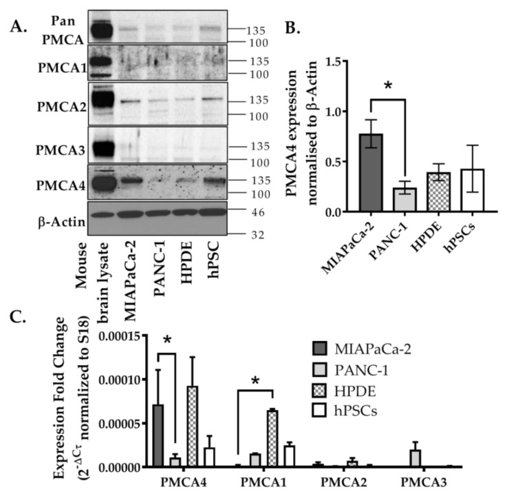
Figure 2.
Expression of PMCA isoforms in multiple pancreatic cell lines. (A) Representative Western immunoblot showing the relative protein expression of total/pan-PMCA and PMCA isoform 1–4 in pancreatic cancer (MIA PaCa-2 and PANC-1) and non-malignant pancreatic cells (human pancreatic ductal epithelial (HPDE) and human pancreatic stellate cells (hPSC)). Mouse brain lysate was used as a positive control for PMCA expressions and β-Actin was used as a protein loading control. (B) PMCA4 protein expression in each cell line was quantified from Western blot bands and normalized to β-Actin housekeeping protein. (C) The relative expressions of ATP2B1–4 (PMCA1–4 mRNA) in each cell line were quantified by RT-qPCR. Data are expressed as relative mRNA expression normalized to corresponding S18 rRNA controls (2−ΔCτ). Statistical comparisons were made using the Kruskal–Wallis test with Dunn’s multiple comparison test and two-way analysis of variance (ANOVA) with Dunnett’s multiple comparison test. Data are expressed as mean ± SEM. (n = 4–5, 4 replicates per treatment condition). * represents statistical significance where p < 0.05.

