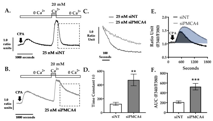
Figure 4.
PMCA4 knockdown in MIA PaCa-2 cells reduces PMCA-mediated Ca2+ clearance. MIA PaCa-2 cells were treated with non-targeting siRNA (siNT) or siRNA targeting PMCA4 mRNA (siPMCA4) for 48–72 hrs prior to performing an in situ Ca2+ clearance assay using fura-2 ratiometric dye. Representative in situ [Ca2+]i traces of MIA PaCa-2 incubated in either (A) 25 nM siNT or (B) 25 nM siPMCA4 are shown. Cells were perfused with Ca2+-free + 1mM EGTA HPSS containing 30 μM CPA (white box) to induce ER intracellular Ca2+ storage depletion. Cells were then treated with HPSS containing 20 mM Ca2+ and 30 μM CPA (grey box) to induce store-operated Ca2+ entry. PMCA-mediated Ca2+ efflux is observed by subsequent removal of extracellular Ca2+ (Ca2+-free HPSS). (C) Expanded time course showing Ca2+ clearance phase (as indicated by dashed box in A.) for siNT vs. siPMCA4 are superimposed. (D) The rate of [Ca2+]i clearance was fitted to a single exponential decay and the time constant (τ) was determined. (E) Expanded time course of the initial CPA-induced increase in [Ca2+]i for siNT (black) vs. siPMCA4 (grey) treated cells are shown. Shaded areas indicate the baseline-subtracted area under the curve (AUC). (F) Data were quantified by measuring the AUC over 1800 sec. Data are shown as mean ± SEM. (n = 5, at least 50 individual cells were analyzed per treatment condition). Comparisons were made between siNT control and siPMCA4 treated cells at matching time points post-drug treatment using Mann–Whitney U-test (Unpaired, non-parametric cumulative distribution test). ** and *** represents statistically significant difference where p < 0.01 and p < 0.001, respectively.

