Figure 7.
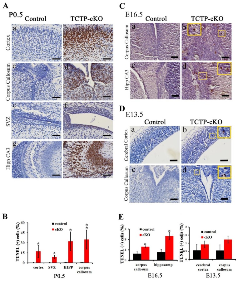
Cell apoptosis was detected in brain sections from control and cKO mice. (A) TUNEL staining was performed on coronal sections of the brain at P0.5, including the (a,b) hippocampus, (c,d) corpus callosum, (e,f) subventricular zone, and (g,h) hippocampus CA3. (B) Bar graph summarizing the quantification of TUNEL-positive (+) cells in (A). (Values are mean ± SEM, Student’s t test, * p < 0.05, ** p < 0.01 compared with control group; n = 3). (C,D) Apoptosis of brain sections from TCTP-cKO and control mice at E16.5 (n = 3) and E13.5 (n = 4) was also detected by TUNEL staining assay. Boxes indicate magnification. (E) Bar graph summarizing quantification of TUNEL-positive cells in (C,D). (Values are mean ± SEM, Student’s t test, * p < 0.05 compared with control group). Hipp, hippocampus; SVZ, subventricular zone. Scale bar: A, C, 40 μm; D, 100 μm.

