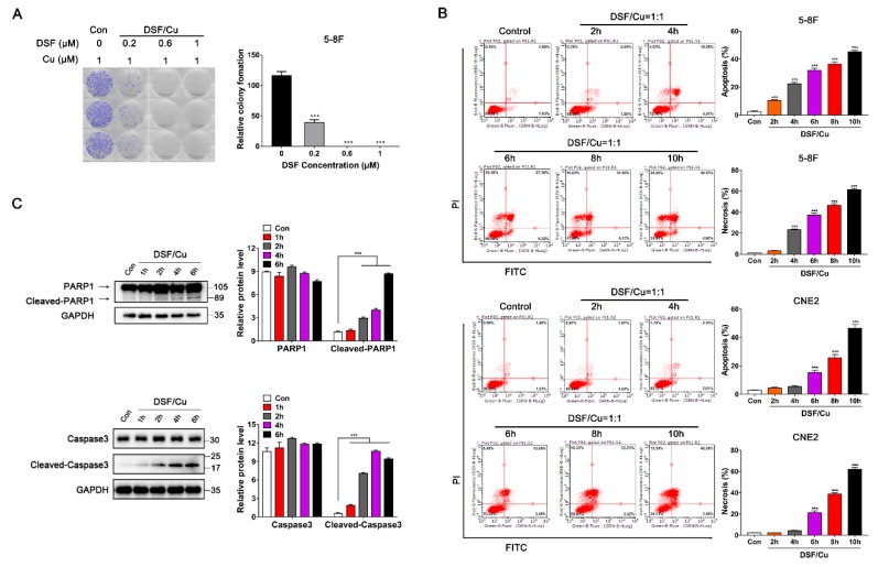
Figure 2.
DSF/Cu promotes the apoptosis and necrosis of nasopharyngeal carcinoma cells. (A) Representative images and quantification of colony formation assay in 6-well plates. 5-8F cells were incubated for 10 days and the medium containing the drug was replaced once. DMSO solvent containing 1 μM Cu was used as a control. Data are shown as means ± SD. *** p < 0.001 vs. control group, n = 3. (B) Flow cytometry with Annexin V/PI double staining proved that DSF/Cu could significantly increase Annexin V+/PI+ cells, and promote the apoptosis and necrosis of 5-8F and CNE2. Data are shown as means ± SD. *** p < 0.001 vs. control group, n = 3. (C) Apoptosis-related protein expressions were detected by Western blot in 5-8F, after being cultured with DSF/Cu (1 μM/1 μM) for different lengths of time. Data are shown as means ± SD. *** p < 0.001, n = 3.

