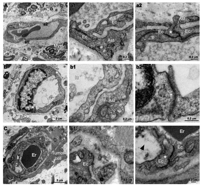
Figure 6.
Transmission electron microscopy (TEM) analysis of human glioma specimens. (A) Representative image of well-structured capillary (Cap)-enclosing erythrocytes (Er) in a low grade astrocytoma (WHO grade II). Tissue and myelinated axonal fibers are still preserved (Myel). Nu, nucleus of endothelial cell. High magnification insets (a1,a2), both indicate “kissing” points of endothelial cell (Ec) tight junctions (asterisk), and astrocytic endfeet (Ast) covering the basal lamina (BL). (B) Panel showing a capillary (Cap) in a region of anaplastic astrocytoma (WHO grade III) without Gd-enhancement. The inter-endothelial junctions are maintained at both sides (asterisks) of endothelial cell nucleus (Nu). Insets (b1,b2) show respectively the luminal part of the vessel (lu) coated by the endothelial wall (Ec), the basal lamina (BL) and the astrocytic endfeet (Ast). (C) panel representative of region of anaplastic astrocytoma (WHO grade III) without Gd-enhancement. Low magnification micrograph showing processes of tumor cell juxtaposed to a blood vessel (Cap) that enclose an erythrocyte (Er). In high magnification insets (c1,c2), both the basal lamina (BL) and the endothelial cell (arrowhead) appear enlarged and swollen, although endothelial cell junctions are still present (asterisk in inset c2).

