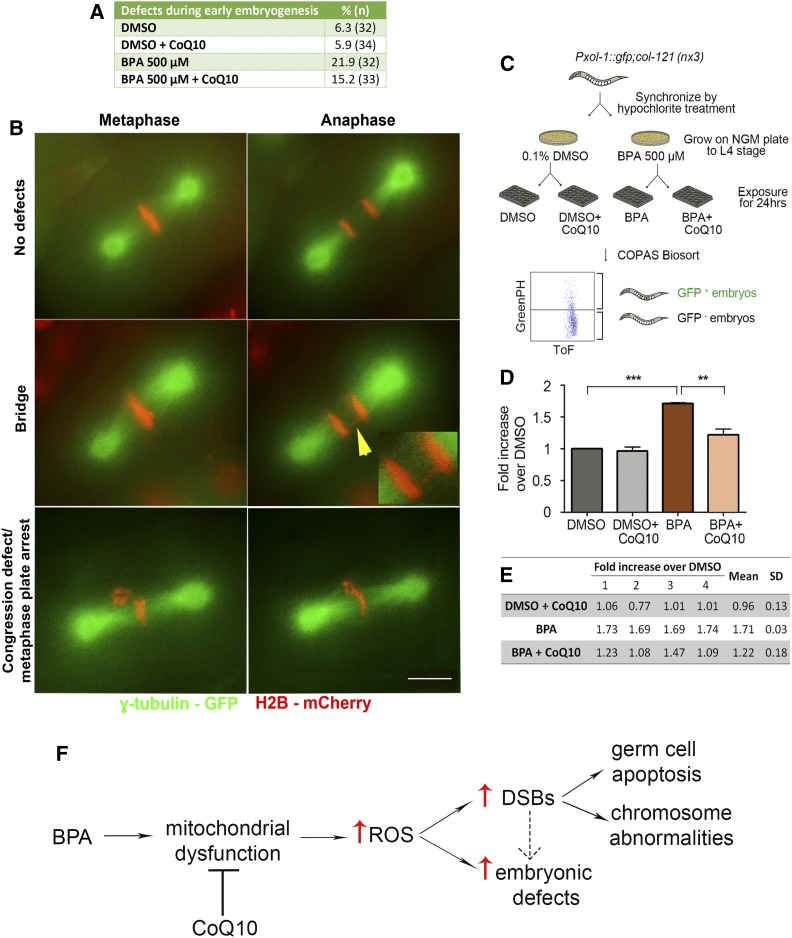
Figure 6.
CoQ10 supplementation rescues BPA-induced defects during the first embryonic division. (A and B) A higher frequency of chromosome segregation defects during the first embryonic cell division is seen in H2B::mCherry; γ-tubulin::GFP; col-121 embryos after treatment with BPA compared to vehicle (DMSO). CoQ10 partially rescues this effect. (A) Quantification of defects detected during the first embryonic cell division. (B) Time-lapse images of the first embryonic division. Shown are examples of normal chromosome alignment at metaphase and segregation at anaphase (top row), with chromosomes in red and spindle microtubules in green. Center and bottom rows show examples of chromatin bridges (yellow arrowhead; insert shows a magnified image of the chromatin bridge), congression defects, and metaphase arrest observed. A Pearson Chi-Square test of independence was calculated for a pairwise comparison of defective and nondefective first embryonic divisions. DMSO used at 0.1%; CoQ10 at 100 µg/ml. Bar, 5 µm. N ≥ 32 per condition. (C–E) High-throughput analysis with the COPAS Biosort shows CoQ10 rescues the elevated chromosome nondisjunction detected during embryogenesis following BPA exposure. (C) Flowchart for high-throughput analysis with the COPAS Biosort. Age-matched L1 stage animals were grown on either DMSO- or BPA-containing NGM plates until reaching the L4 stage. L4 stage animals from DMSO plates were dispensed into 24-well plates (300 worms/well) containing OP50 E. coli (OD600 = 24) in M9 buffer and either DMSO alone or DMSO with CoQ10, and L4 stage animals from BPA plates were dispensed into 24-well plates with either BPA or BPA with CoQ10. After a 24-hr incubation at 20°, worms were thoroughly washed with M9 and sorted with the COPAS Biosort based on fluorescence intensity. Time-of-flight (Tof) and GFP peak height were used as reading parameters. (D) Levels of GFP+ embryos (resulting from X chromosome nondisjunction) detected in the uterus of BPA-treated worms are significantly higher relative to DMSO (***P < 0.0001) and CoQ10 treatment results in a significant reduction in the elevation induced by BPA (**P = 0.0021). DMSO used at 0.1%; BPA at 500 µM; CoQ10 at 100 µg/ml. Statistical comparisons were performed using the unpaired two-tailed t-test, 95% C.I. Error bars represent SEM. (E) Readouts obtained with the COPAS Biosort for the indicated chemicals. Shown is the fold increase over DMSO alone calculated for each biological repeat (labeled as 1 through 4). SD, standard deviation. (F) Model for how CoQ10 restores fertility by rescuing BPA-induced oxidative DNA damage in the C. elegans germline. BPA exposure may lead to mitochondrial dysfunction in the germline resulting in increased reactive oxygen species (ROS) that in turn may account, at least in part, for the increased levels of DNA double-strand breaks (DSBs) and the embryonic defects observed. Unrepaired or aberrantly repaired meiotic DSBs lead to the activation of a late pachytene DNA damage checkpoint resulting in increased germ cell apoptosis, the increased chromosome abnormalities detected in oocytes at late diakinesis, and some of the observed embryonic defects (the latter is depicted by a dashed arrow, given that a direct effect on embryos may also be possible). Only the pathway/mode of action directly tested in this study is depicted, but other studies have shown additional effects such as alterations in gene expression (Allard and Colaiacovo 2010; Chen et al. 2016).

