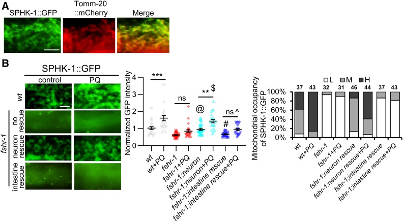
Figure 4.
FSHR-1 regulates mitochondrial association of SPHK-1. (A) High-magnification images of SPHK-1::GFP (left), TOMM-20::GFP (middle), and merge (right) in wt animals coexpressing Pges-1::sphk-1::gfp and Pges-1::tomm-20::mCherry transgenes. (B) Left: representative images of the indicated mutants expressing intestinal SPHK-1::GFP in the absence or presence of PQ for 24 hr. fshr-1 neuron and intestine rescue denote transgenic fshr-1 mutants expressing fshr-1 cDNA under the pan-neural promoter rab-3 or the intestinal promoter ges-1, respectively. Middle: average fluorescence intensities in the indicated strains expressing SPHK-1::GFP are quantified. Right: mitochondrial occupancy of indicated transgenes is quantified in the absence or presence of PQ. H, M, or L indicates the percentage of animals showing mitochondrially localized SPHK-1::GFP in more than two-thirds of the intestine (H), one-half of the intestine (M), or only in a few intestinal cells (L). Bar represents 2 µm. One-way ANOVA with Bonferroni post-test **P < 0.01; ***P < 0.001; @P > 0.05 compared to wt; #P > 0.05 compared to fshr-1 mutants; $P > 0.05 compared to wt + PQ; and ^P > 0.05 compared to fshr-1 + PQ. cDNA, complementary DNA; ns, not statistically significant; PQ, paraquat; wt, wild-type.

