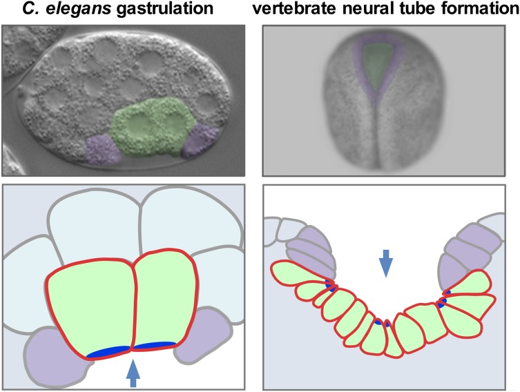
Figure 1.
C. elegans gastrulation and vertebrate (Xenopus) neural tube formation. Photos (A and B) and cross-section diagrams (C and D)) of C. elegans and Xenopus embryos, with internalizing cells (just the endodermal precursor cells in C. elegans and neural plate cells in Xenopus) colored green. Apical neighbors that are represented in the cross-section diagrams are colored purple. Arrows mark the direction of internalization in the views shown. Bright blue represents the apical parts of cells that undergo apical constriction. Modified from Sullivan-Brown et al. (2016).

