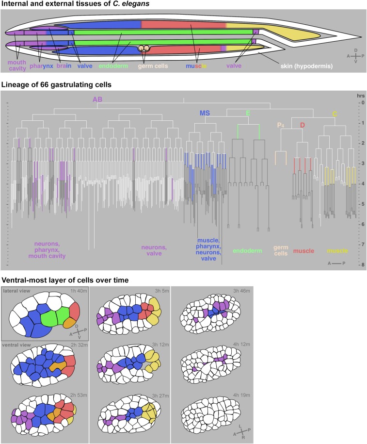
Figure 2.
Movement of C. elegans cells from the surface to the interior during gastrulation. Diagram of internal and external tissues of C. elegans at the end of embryogenesis (top). Lineage with all 66 cells identified to gastrulate marked in colors and their cell fates indicated (middle). Horizontal lines are cell divisions and vertical lines are cells. White lines are exterior cells; colored lines are gastrulating cells; and the progeny of gastrulating cells, most or all of which are interior, are in gray. First nine rounds of embryonic cell divisions are drawn to a total of 409 cells, based on cell division timing data from WormBase release WS170. Tracings of one plane of an embryo over time depicting the ventral surface cells at all but the first timepoint (bottom). Times are marked in hours and minutes after the one-cell-stage division. Some anterior AB lineage-derived cells are not shown because they internalize from the side not shown in the embryos at the bottom. Progenitors of cells that will internalize are also colored, except in lineages that produce some external cells (AB and C lineages), which for clarity are left white until the last or second-to-last cell cycle before internalization in the AB lineage. Gray letters in lower right of some illustrations indicate axes: A (anterior) and P (posterior), D (dorsal) and V (ventral), and L (left) and R (right). White represents cells on the exterior and cells that internalize are marked in color. For movie, see https://youtu.be/BaV63cLO1Tg. Modified from Harrell and Goldstein (2011), which can be referred to for more detailed information.

