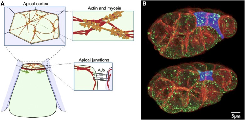
Figure 5.
Actomyosin, apical junctions, and apical constriction. (A) Components involved in apical constriction include F-actin (red) and nonmuscle myosin II (orange), which form contractile networks. Network is shown at sparse density for purpose of illustration. Shrinkage of the apical cortex (green arrows) is driven by contraction of apical actin–myosin networks linked to apical adherens junctions (AJs, gray), resulting in tissue shape changes. Modified from Martin and Goldstein (2014). (B) Bessel beam structured plane illumination images of an embryo at two timepoints (5 min, 40-sec apart) during endodermal precursor cell internalization, with membranes in red, myosin in green, and apical surfaces of endodermal precursor cells false-colored blue. From Roh-Johnson et al. (2012).

