Figure 5.
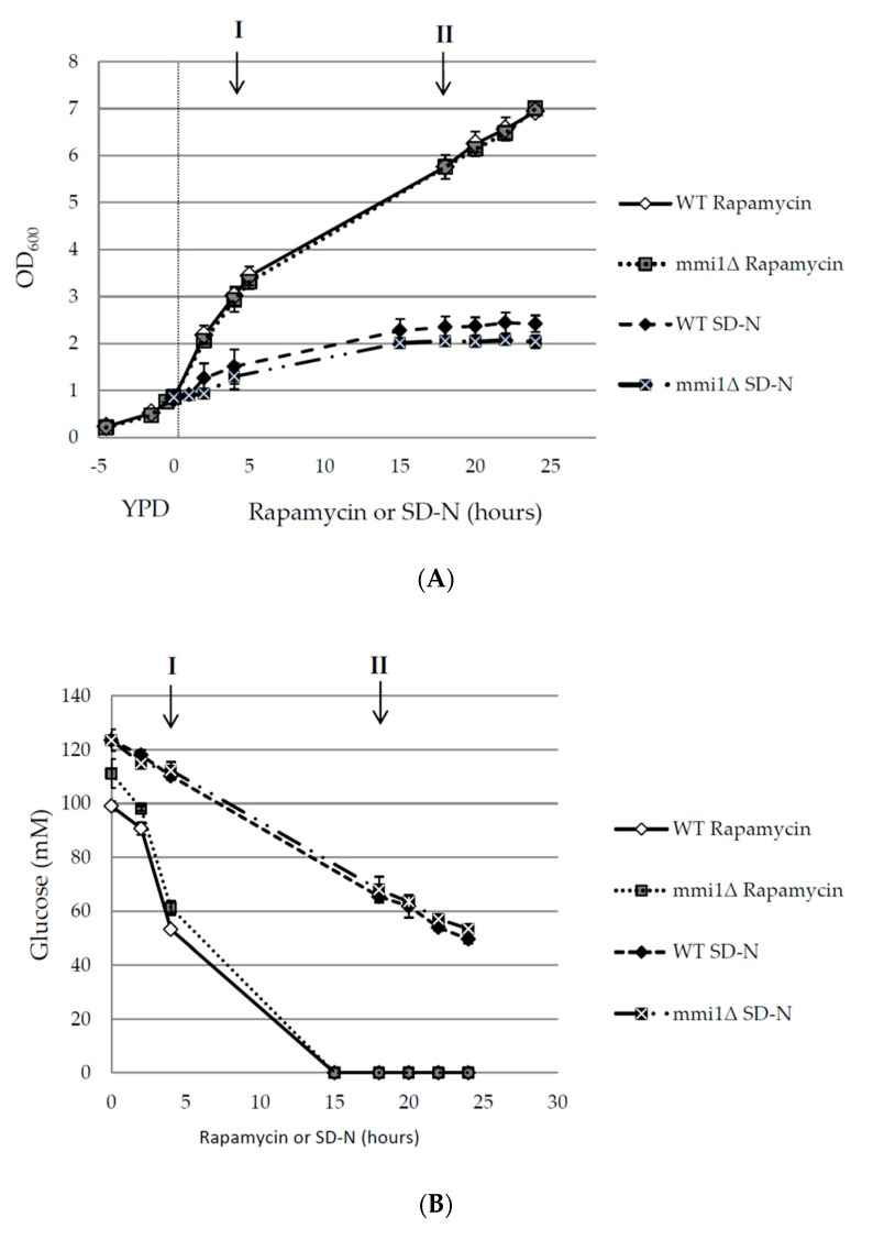
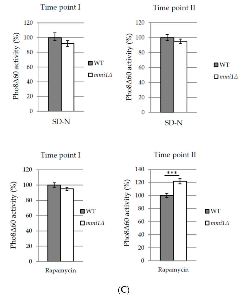
Increased rapamycin-induced autophagy in mmi1Δ strain occurs after glucose exhaustion. (A) Exponentially growing WT and mmi1∆ cells expressing Pho8Δ60 (OD600 ≈ 0.8) in the YPD medium were either shifted to the SD-N medium or treated with rapamycin (200 nM). Optical density at 600 nm was measured at indicated time points. Results are presented as means ± SD of three independent experiments performed in duplicates (n = 6). (B) Concentration of glucose in the media as shown in A was measured after the addition of rapamycin (200 nM) to WT and mmi1∆ strains or after the shift of the strains to SD-N media. Results are means of two independent experiments performed in triplicates (n = 6). (C) Pho8Δ60 assay was measured in time points I and II as indicated in A, and B. Point I represents the exponential growth phase where glucose is still present in the medium. Point II represents the post-diauxic phase where glucose is already exhausted. Results are normalized to the WT strain (100%) and represent means ± SD from two independent experiments performed in duplicates (n = 4). The statistical evaluation was performed by using two way analysis of variance (ANOVA). *** p < 0.001.


