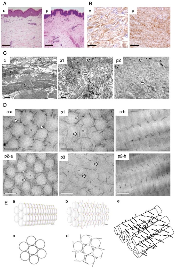
Figure 4.
Skin pathology of mcEDS-CHST14. (A) Light microscopy (hematoxylin and eosin staining). In the skin specimen from a patient with heterozygous variants Pro281Leu/ Cys289Ser (panel p), fine collagen fibers are present predominantly in the reticular to papillary dermis with marked reduction of thick collagen bundles; thick collagen bundles are observed in a skin specimen from a healthy control volunteer (panel c) [6]. (B) Immunohistochemical staining for decorin core protein. Decorin core protein is present on collagen fibers in thick bundles in a skin specimen from a healthy control volunteer (panel c), but on thin and filamentous collagen fibers without clear boundaries in a skin specimen from a patient with heterozygous variants Pro281Leu/ Cys289Ser (panel p) [31] (C) Transmission electron microscopy. Collagen fibrils are regularly and tightly assembled in a skin specimen from a healthy control volunteer (panel c), but are dispersed in the papillary to reticular dermis in skin specimens from a patient with heterozygous variants Pro281Leu/ Tyr293Cys (panel p1) and a patient with a novel homozygous variant (panel p2) [31]. (D) Transmission electron microscopy-based cupromeronic blue staining. GAG chains are curved and maintain close contact with attached collagen fibrils in the skin specimens from a healthy control volunteer (panels c-a, c-b); conversely, they are linear and stretch from the outer surface of collagen fibrils to adjacent fibrils in skin specimens from a patient with heterozygous variants Pro281Leu/ Tyr293Cys (panel p1), a patient with a novel homozygous variant (panels p2a and p2b) and another patient with heterozygous variants Pro281Leu/ Tyr293Cys (panel p3) [31]. (E) Schematic representations of collagen fibrils and GAG chains. Decorin core protein binds to D bands of collagen fibrils both in normal skin and in affected skin. GAG chains composed of DS adhere to collagen fibrils along D bands, beginning from the core protein (panels a and c), whereas GAG chains composed of CS extend linearly and perpendicularly to collagen fibrils from the core protein (panels b and d) [31]. Putative spatial disorganization of collagen fibril networks in the skin of patients (panel e) (A, reproduced from Miyake et al. Hum. Mutat. 2010, 31, 1233–1239, with permission from Wiley-Liss, Inc.; B–E, reproduced from Hirose et al. Biochim. Biophys. Acta Gen. Subj. 2019, 1863, 623–631, with permission from Elsevier, Inc.).

