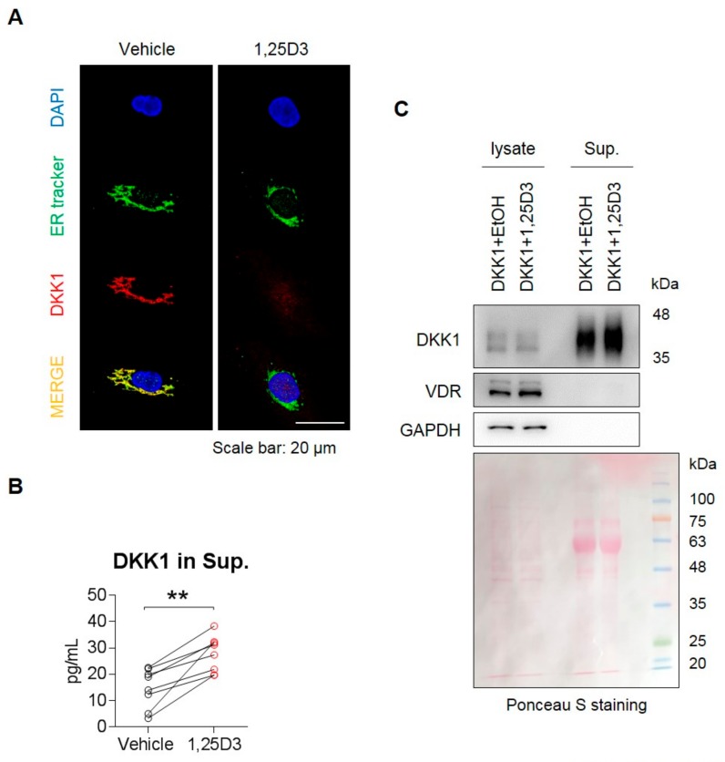
Figure 4.
1,25D3 stimulates secretion of DKK1 protein in osteoblasts. (A) Osteoblasts were treated with 20 nM 1,25D3 for 24 h and stained with ER tracker (green), anti-DKK1 (red), and DAPI (blue). Scale bar is 20 μm. Representative data are shown (n = 3). (B) Osteoblasts were treated with 20 nM 1,25D3 for 24 h, then the culture supernatant was collected and secreted DKK1 protein was measured in the supernatant using ELISA. ** p < 0.01 (mean ± SD; n = 6). (C) Osteoblasts were transfected with 2 μg DKK1 plasmid for 48 h, and then treated with 1,25D3 for 24 h. Proteins secreted in the cell supernatant by 1,25D3 stimulation were collected, precipitated by trichloroacetic acid (TCA) and detected by SDS-PAGE/immunoblotting. Ponceau S staining was used as culture supernatant controls. Representative data are shown (n = 3).

