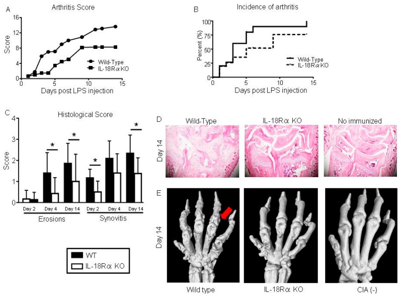
Figure 2.
Inhibition of collagen-induced arthritis (CIA) in IL-18Rα knock-out (KO) mice. (A) Disease severity was assessed by visual inspection and scored on a scale of 0–4 per paw, 2×/week. The arthritis score (0–16) of CIA in the wild-type (WT; n = 30) and IL-18Rα KO mice (n = 29) after lipopolysaccharide (LPS) injection was calculated for each treatment group, and differences between groups were analyzed by the Mann-Whitney test. (B) The incidence of arthritis from grade 1 in each mouse is presented as Kaplan-Meier curves and was analyzed by log-rank test. (C) Microscopic synovitis and erosions on bone and cartilage were assessed the histological score in wrist sections stained with hematoxylin and eosin (H&E) from WT and IL-18Rα KO mice with CIA on day 2 (n = 12 and n = 8), 4 (n = 10 and n = 15), and 14 (n = 8 and n = 6). (D) Representative histological H&E images of mouse wrist joints in the WT, IL-18Rα KO, and non-immunized mice on day 14. (E) Visualization of CIA-induced joint damage in the wrist joints by micro-CT on day 14. Micro-CT imaging of a representative wrist from an IL-18Rα KO mouse with CIA shows normalized joint architecture and the prevention of bone destruction (red arrow). The data are mean ± SEM. * p < 0.05, ** p < 0.01, WT vs. IL-18Rα KO mice. Original magnification, ×40.

