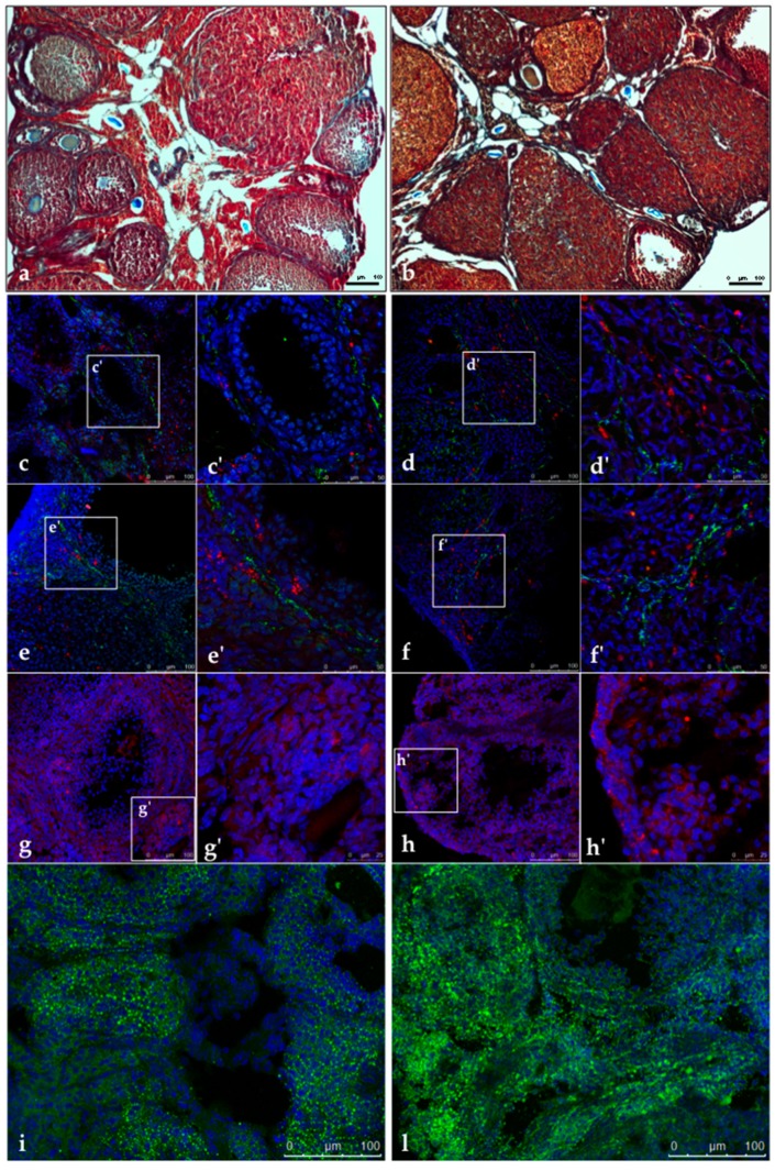
Figure 2.
Representative images of trichrome staining in CTRL (a) and DHEA (b) mice. (c–f) Immunolocalization of Von Willebrand Factor (vWF) (green) and alpha smooth muscle actin (α-SMA) (red) in control (c,c’ and e,e’) and DHEA (d,d’ and f,f’) ovarian sections. (g–h) Immunolocalization of 17 beta-hydroxysteroid dehydrogenase type 4 (17β-HSD4) (red) in control (g) and DHEA (h) ovarian sections. Staining of lipid droplets by BODIPY 493/503 (green) in control (i) and DHEA (l) ovarian section. (c–l) DNA is stained by DAPI (blue). Three mice per experimental group were employed. Experiments were done in triplicate.

