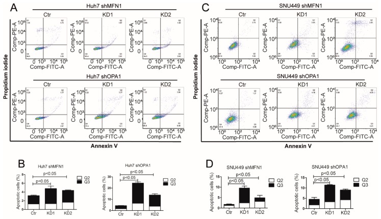
Figure 5.
Mitochondrial fusion dysfunction inhibited tumor growth through cell apoptosis induction (A,B) Apoptotic cells were quantified by flow cytometry using Annexin V and propidium iodide co-staining in Huh7 shOPA1 and Huh7 shMFN1 cells (n = 3). (C,D) Apoptotic cells were quantified by flow cytometry using Annexin V and propidium iodide co-staining in SNU449 shOPA1 and SNU449 shMFN1 cells (n = 3). Histograms show means ± SEM with p value derived by the Mann Whitney test.

