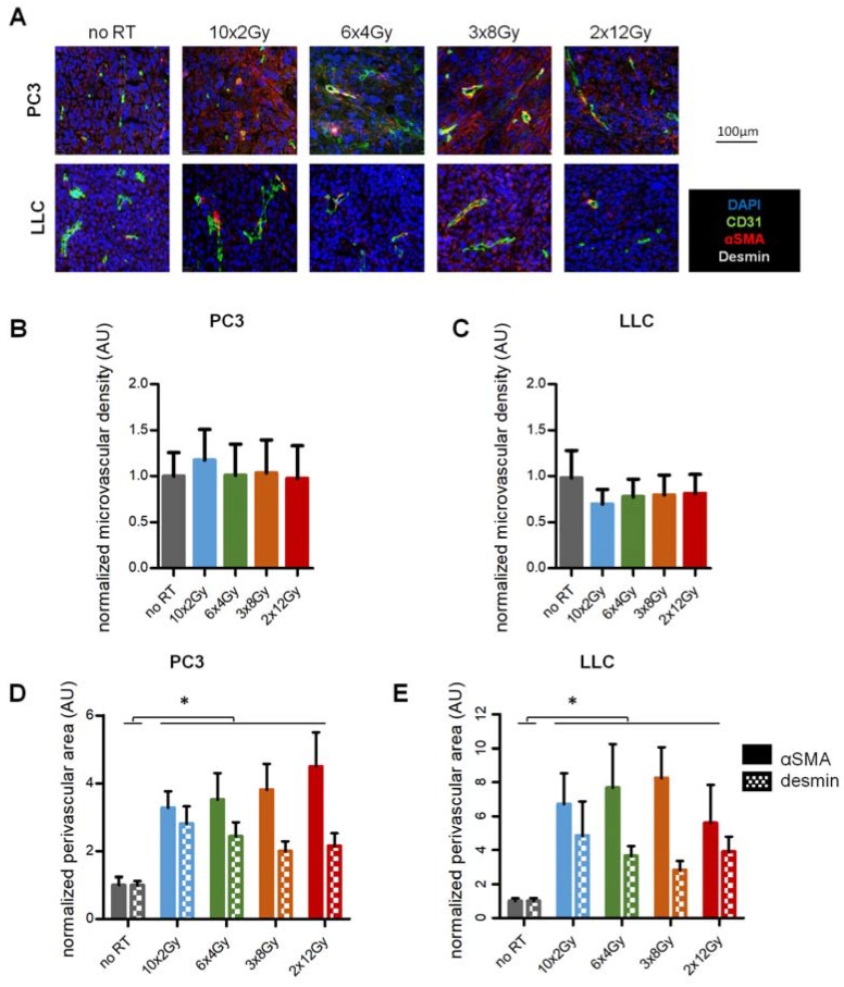
Figure 2.
Tumor vascular phenotype in response to RT fractionation schedule. (A) Immunohistological staining for pericyte coverage (αSMA, desmin) around tumor vessels (CD31) two weeks after RT initiation in PC3 and LLC tumors. (B,C) Quantification of vessel density in PC3 (B) and LLC (C) tumors at day 15. (D,E) Quantification of vascular mural coverage (αSMA: plain, desmin: squared) in PC3 (D) and LLC (E) tumors at day 15. Images and analysis represent two independent experiments with a total ≥18 tumors per point. * indicates p < 0.05.

