Figure 3.
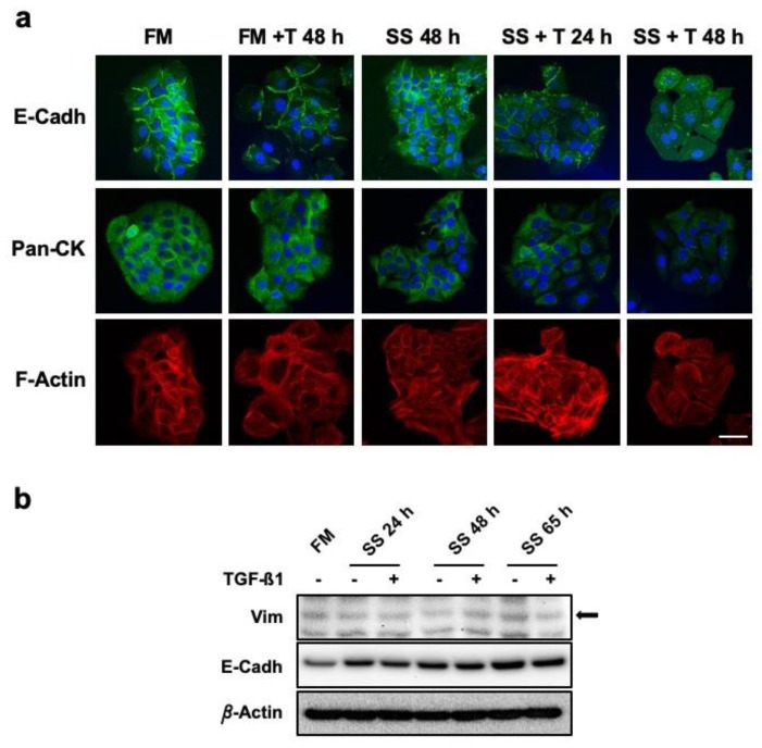
HaCaT keratinocytes do not experience EMT in response to continuous TGF-β1 exposure. (a) Confocal microscopy studies on the expression and localization of structural markers E-Cadherin (E-Cadh), Citokeratins (Pan-Ck), and actin filaments (F-Actin) with, or without, TGF-β1. Scale bar: 50 µm. (b) Protein levels of Vimentin (Vim) and E-Cadherin (E-Cadh) were assessed by Western Blot. Total protein extracts from HaCaT cells cultured for the indicated times in SS conditions, with or without TGF-β1, were compared to control sample (FM). β-Actin was used as protein loading control. Arrow indicates the correct identity of protein bands. Representative images of at least three independent experiments are shown. FM: full medium; SS: serum starvation; + T24 h, + T48 h: time (hours) of continuous TGF-β1 stimulation.

