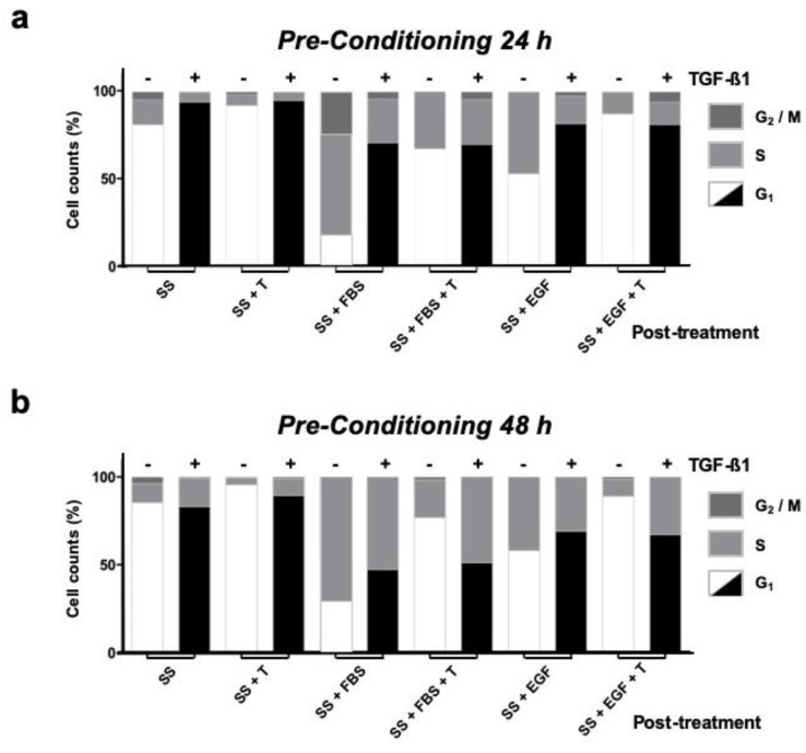
Figure 5.
HaCaT cells under continuous TGF-β1 treatment show impaired TGF-β1 restraint with serum or EGF stimuli. Keratinocytes in serum deprived medium were conditioned by continued exposure to TGF-β1 [+] for either 24 h (a) or 48 h (b). Untreated cells (-)were used as reference controls. Response to post-treatment stimulus was determined by flow cytometry. Black G1 columns represent cells maintained in continuous serum deprived conditions with TGF-β1; white G1 columns represent control cells maintained in serum deprived conditions without TGF-β1. SS: Serum starvation medium; + T: TGF-β1 inoculation; + FBS: Foetal bovine serum supplementation; EGF: Epidermal growth factor inoculation. Representative data from at least three independent experiments are shown.

