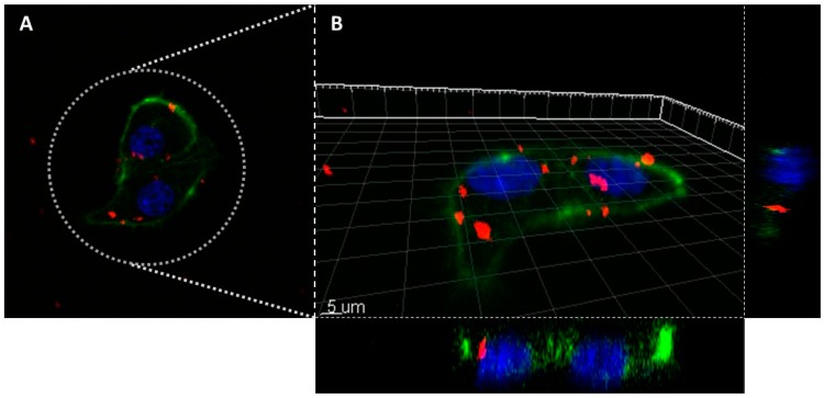
Figure 8.
Confocal laser scanning microscope images of the MDA-MB-231 cells incubated overnight with the nanomaterial AX-3 (A): 2D image; (B): 3D reconstruction). Colors legend: MSN nanoparticles are in red (Alexa Fluor 647, Ex.650/Em.665 nm), cellular nucleus are in blue (DAPI staining, Ex.358/Em.461) and actin filaments are in green (Phalloidin staining, Ex.495/Em.519 nm).

