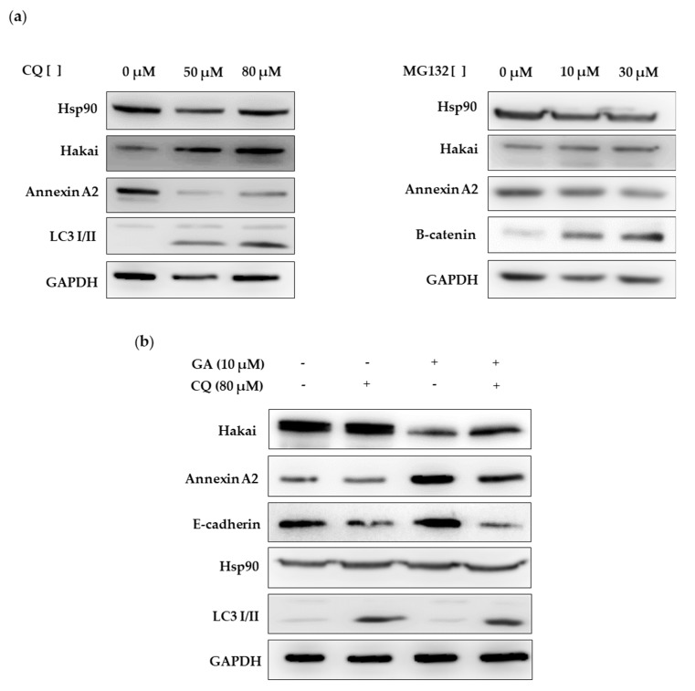
Figure 7.
Geldanamycin increases Hakai protein degradation in a lysosome-dependent manner: (a) HEK293T cells were treated with chloroquine (left panel) and with MG132 (right panel) for 24 h at the indicated concentrations. Cell lysates were collected and evaluated by western blot analyses with Hakai and Annexin A2 antibodies. (b) HEK293T cells were transiently transfected with pcDNA-Flag-Hakai (4 µg), pBSSR-HA-Ubiquitin (3 µg) and pSG-v-Src (3 µg) for 48 h. The day after transfection, cells were treated with chloroquine and geldanamycin at the indicated concentrations for 24 h. Cell lysates were collected and protein expression was evaluated by western blot analyses using the indicated antibodies. Chloroquine treatment-induced Hakai protein levels recovery during geldanamycin treatment. LC3 I/II levels was used as a positive control in chloroquine treatment and β-catenin as positive control in MG132 treatment.

