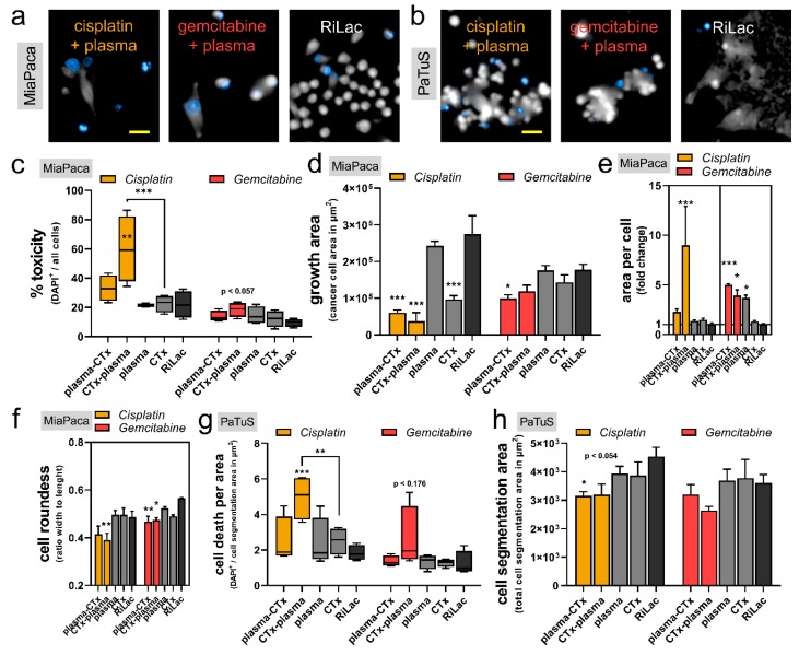
Figure 4.
A combination of cisplatin and gemcitabine with physical plasma-conditioned Ringer’s lactate was toxic, reduced the cancer cells’ growth, and altered their morphology. (a) Representative images with the DAPI fluorescence channel and digital phase contrast of MiaPaca cells (scale-bar = 50 µm); (b) representative images with the DAPI fluorescence channel and digital phase contrast of PaTuS cell (scale-bar = 50 µm); (c–f) algorithm-based quantification of high-content imaging experiments showing (c) the toxicity (% DAPI+ events on all events), (d) the growth area (area of the pseudo-cytosolic digital phase contrast), (e) the area per cell, and (f) and roundness of MiaPaca pancreatic cancer cells; (g,h) algorithm-based quantification of (g) cell death (DAPI+ events per area) and (h) cell segmentation area of treated PaTuS cells. Imaging was performed at 48 h post initial exposure to the treatment liquids. Data are representative out of three independent experiments and are presented as (c,g) boxplot with their median ± min and max, or (e–h) as mean + SEM. Statistical significance was calculated utilizing ANOVA.

