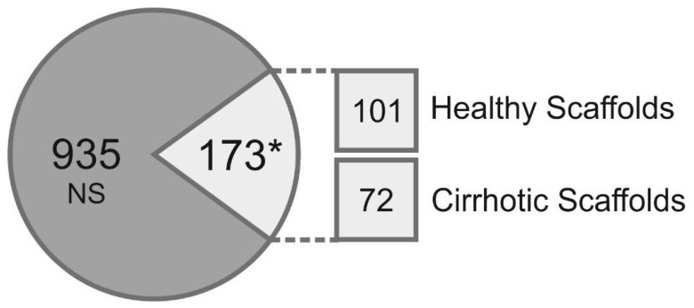
Figure 3.
Proteomic analysis of healthy liver 3D scaffolds and cirrhotic liver 3D scaffolds. The composition of the ECM in decellularized healthy and cirrhotic liver scaffolds was qualitatively and quantitatively investigated by a label free proteomic analysis (n = 3, three biological repeats, processed in triplicate for each condition). A relative quantitative analysis was performed on 1108 proteins showing 173 proteins significantly changed (* p < 0.05) between healthy scaffold and cirrhotic 3D scaffold ECM. In 3D healthy scaffolds, 101 proteins were overexpressed, whereas 72 proteins were significantly changed in decellularized 3D cirrhotic liver scaffolds.

