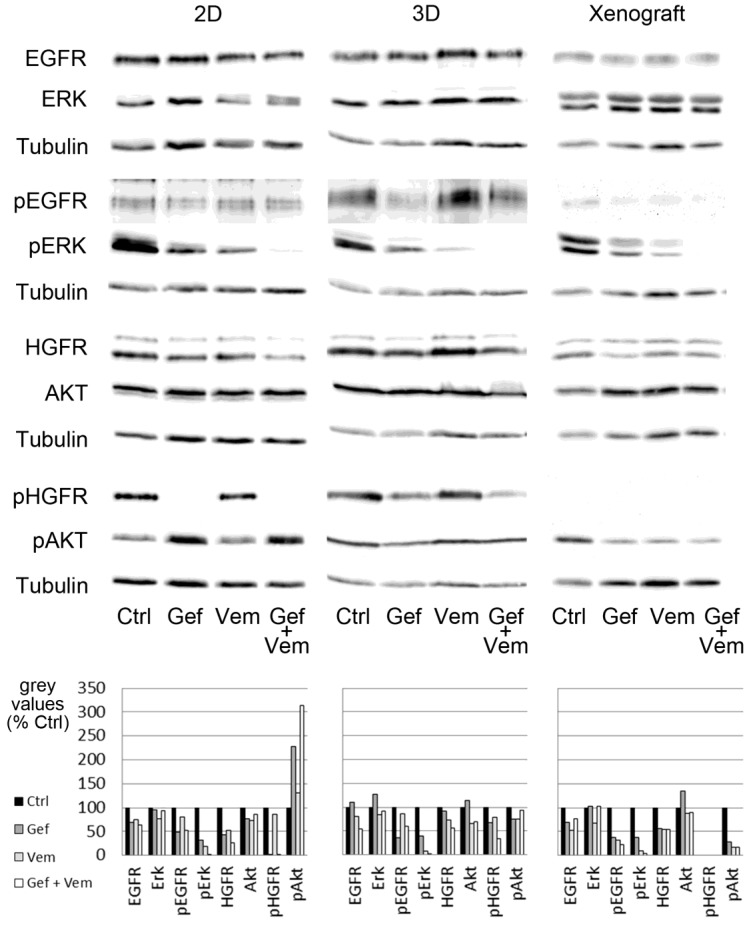
Figure 5.
Signaling in HROC87 cells upon treatment with vemurafenib and/or gefitinib. Shown is the Western blot analysis of the phosphorylation status of molecules involved in signal transduction for proliferation (ERK) and apoptosis (AKT) and important receptors in HROC87 cells cultured under 2D and 3D conditions as well as in matching xenografts. In 2D, 3D and PDX, ERK is partly inhibited by gefitinib and vemurafenib in monotherapy, but nearly completely inhibited by the combination-therapy. In contrast to 2D, AKT phosphorylation show no clear upregulation under 3D culture conditions. HGFR could not be activated in the PDX at all. Only in the 2D model, gefitinib and the combination-therapy results in a strong inactivation of the HGFR. Ctrl: untreated control, Gef: gefitinib, Vem: vemurafenib, Gef + Vem: combination-therapy of gefitinib and vemurafenib. Grey values of densitometric analysis of the representative blots are shown in the diagrams (% of Ctrl). Blots with weight markers are shown in Figure A4, Figure A5 and Figure A6. Densitometric analyses of grey values from all 4 to 6 experiments (Figure A7) as well as from the representative blot depicted here (Table A1) are shown in the appendix.

