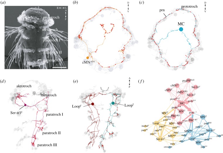
Figure 4.
The ciliomotor circuit of the Platynereis larva. (a) SEM of a Platynereis nectochaete (3 days old) larva with ciliary bands labelled. Scale bar 50 µm. (b) serial scanning transmission electron microscopy (ssTEM)-based reconstructions of one of three catecholaminergic neurons (anterior view) and (c) of the closure-inducing cholinergic MC neuron (anterior view) in the Platynereis ciliomotor circuit. Ciliated cells are shown in grey. (d) Reconstruction of the serotonergic Ser-tr1 and (e) cholinergic Loop ciliomotor neurons (ventral views). (f) Synaptic connectivity graph of all ciliomotor neurons and ciliary band cells.

